Anti-AURKA antibody [A4-8D]
-
概述
- 产品描述Aurora related kinase-1 (ARK-1, STK15, Aurora2, Aik1) and -2 (ARK-2, STK12, Aurora1) are centrosome-associated serine/threonine kinases that regulate centrosome separation, bipolar spindle assembly and chromosome segregation during mitosis. ARK-1 and -2 are expressed in the nucleus and localize to distinct portions of mitotic machinery such as the centrosome, spindle poles (ARK-1) and midbody (ARK-2) during mitosis. ARK-1 and -2 transcripts are present at high levels in human thymus and fetal liver. ARK-1 protein has elevated expression in colon carcinoma lines (HT-29, SNU-C2B, COLO 205, SW480, 837 and 948) and accumulates during metaphase in HeLa cells. ARK-2 protein levels are maximal during both S and G2/M phases, whereas ARK-1 protein is degraded after G2/M via the ubiquitin-proteasome pathway. ARK-2 has a unique genetic loci relative to ARK-1, suggesting that these two kinases, with oncogenic potential, have different roles in cell cycle progression.
- 产品名称Anti-AURKA antibody [A4-8D]
- 分子量46 kDa
- 种属反应性Human
- 验证应用WB,ICC,IHC-P,FC
- 抗体类型小鼠单抗
- 免疫原Recombinant protein
- 偶联Non-conjugated
-
性能
- 形态Liquid
- 浓度2 mg/mL.
- 存放说明Store at +4℃ after thawing. Aliquot store at -20℃ or -80℃. Avoid repeated freeze / thaw cycles.
- 存储缓冲液1*TBS (pH7.4), 1%BSA, 40%Glycerol. Preservative: 0.05% Sodium Azide.
- 亚型IgG2b
- 纯化方式Protein A purified.
- 亚细胞定位Cell membrane. Endosome membrane. Lysosome membrane
- 其它名称
- AIK antibody
- ARK-1 antibody
- ARK1 antibody
more
-
应用
WB: 1:500-1:2,000
ICC: 1:50-1:200
IHC-P: 1:50-1:200
FC: 1:50-1:100
-
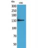
Fig1: Western blot analysis of Aurora A on human Aurora A recombinant protein using anti- Aurora A antibody at 1/1,000 dilution.
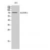
Fig2: Western blot analysis of Aurora A on HEK293 (1) and Aurora A -hIgGFc transfected HEK293 (2) cell lysate using anti- Aurora A antibody at 1/1,000 dilution.
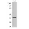
Fig3: Western blot analysis of Aurora A on different cell lysate using anti- Aurora A antibody at 1/1,000 dilution.
Positive control: Line1: HEK293 Line2: MCF-7 Line3: Hela
Fig4: ICC staining Aurora A (green) and Actin filaments (red) in Hela cells. The nuclear counter stain is DAPI (blue). Cells were fixed in paraformaldehyde, permeabilised with 0.25% Triton X100/PBS.
Fig5: ICC staining Aurora A (green) and Actin filaments (red) in SMMC-7721 cells. The nuclear counter stain is DAPI (blue). Cells were fixed in paraformaldehyde, permeabilised with 0.25% Triton X100/PBS.
Fig6: Immunohistochemical analysis of paraffin-embedded human cervical cancer tissue using anti- Aurora A antibody. Counter stained with hematoxylin.
Fig7: Immunohistochemical analysis of paraffin-embedded human rectum cancer tissue using anti- Aurora A antibody. Counter stained with hematoxylin.
Fig8: Flow cytometric analysis of Hela cells with Aurora A antibody at 1/100 dilution (green) compared with an unlabelled control (cells without incubation with primary antibody; red).
特别提示:本公司的所有产品仅可用于科研实验,严禁用于临床医疗及其他非科研用途!

![Anti-AURKA antibody [A4-8D]](images/202012/goods_img/92747_G_1607938545949.jpg)