Anti-MMP9 antibody [10A1]
-
概述
- 产品描述The matrix metalloproteinases (MMP) are a family of peptidase enzymes responsible for the degradation of extracellular matrix components, including collagen, gelatin, fibronectin, laminin and proteoglycan. Transcription of MMP genes is differentially activated by phorbol ester, lipopolysaccharide (LPS) or staphylococcal enterotoxin B (SEB). MMP catalysis requires both calcium and zinc. MMP-9 (also designated 92 kDa type IV collagenase or gelatinase B) has been shown to degrade bone collagens in concert with MMP-1 (also designated interstitial collagenase, fibroblast collagenase or collagenase-1), and cysteine proteases and may play a role in bone osteoclastic resorption. MMP-1 is downregulated by p53, and abnormality of p53 expression may contribute to joint degradation in rheumatoid arthritis by regulating MMP-1 expression.
- 产品名称Anti-MMP9 antibody [10A1]
- 分子量64/67/78 kDa
- 种属反应性Human
- 验证应用WB,ICC,IHC-P,FC
- 抗体类型小鼠单抗
- 免疫原Recombinant protein with Human MMP-9 aa 270-400.
- 偶联Non-conjugated
-
性能
- 形态Liquid
- 浓度2 mg/mL.
- 存放说明Store at +4℃ after thawing. Aliquot store at -20℃. Avoid repeated freeze / thaw cycles.
- 存储缓冲液1*PBS (pH7.4), 0.2% BSA, 50% Glycerol. Preservative: 0.05% Sodium Azide.
- 亚型IgG1
- 纯化方式Protein G purified.
- 亚细胞定位Extracellular matrix. Secreted.
- 其它名称
- 82 kDa matrix metalloproteinase-9 antibody
- 92 kDa gelatinase antibody
- 92 kDa type IV collagenase antibody
more
-
应用
WB:1:500
ICC:1:50-1:200
IHC-P:1:50-1:200
FC:1:50-1:100
-
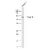
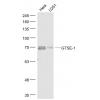
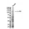
Fig1: Western blot analysis of MMP-9 on MCF-7 cell lysate. Proteins were transferred to a PVDF membrane and blocked with 5% BSA in PBS for 1 hour at room temperature. The primary antibody was used at a 1:500 dilution in 5% BSA at room temperature for 2 hours. Goat Anti-Mouse IgG - HRP Secondary Antibody (HA1006) at 1:5,000 dilution was used for 1 hour at room temperature.
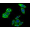
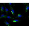
Fig2: ICC staining MMP9 in PANC-1 cells (green). Formalin fixed cells were permeabilized with 0.1% Triton X-100 in TBS for 10 minutes at room temperature and blocked with 1% Blocker BSA for 15 minutes at room temperature. Cells were probed with MMP9 antibody at a dilution of 1:100 for 1 hour at room temperature, washed with PBS. Alexa Fluorc™ 488 Goat anti-Mouse IgG was used as the secondary antibody at 1/100 dilution. The nuclear counter stain is DAPI (blue).
Fig3: ICC staining MMP9 in SH-SY5Y cells (green). Formalin fixed cells were permeabilized with 0.1% Triton X-100 in TBS for 10 minutes at room temperature and blocked with 1% Blocker BSA for 15 minutes at room temperature. Cells were probed with MMP9 antibody at a dilution of 1:100 for 1 hour at room temperature, washed with PBS. Alexa Fluorc™ 488 Goat anti-Mouse IgG was used as the secondary antibody at 1/100 dilution. The nuclear counter stain is DAPI (blue).
Fig4: Immunohistochemical analysis of paraffin-embedded human tonsil tissue using anti-MMP9 antibody. The section was pre-treated using heat mediated antigen retrieval with Tris-EDTA buffer (pH 8.0-8.4) for 20 minutes.The tissues were blocked in 5% BSA for 30 minutes at room temperature, washed with ddH2O and PBS, and then probed with the antibody at 1/50 dilution, for 30 minutes at room temperature and detected using an HRP conjugated compact polymer system. DAB was used as the chrogen. Counter stained with hematoxylin and mounted with DPX.
Fig5: Immunohistochemical analysis of paraffin-embedded human lung cancer tissue using anti-MMP9 antibody. The section was pre-treated using heat mediated antigen retrieval with Tris-EDTA buffer (pH 8.0-8.4) for 20 minutes.The tissues were blocked in 5% BSA for 30 minutes at room temperature, washed with ddH2O and PBS, and then probed with the antibody at 1/200 dilution, for 30 minutes at room temperature and detected using an HRP conjugated compact polymer system. DAB was used as the chrogen. Counter stained with hematoxylin and mounted with DPX.
Fig6: Immunohistochemical analysis of paraffin-embedded human spleen tissue using anti-MMP9 antibody. The section was pre-treated using heat mediated antigen retrieval with sodium citrate buffer (pH 6.0) for 20 minutes. The tissues were blocked in 5% BSA for 30 minutes at room temperature, washed with ddH2O and PBS, and then probed with the antibody at 1/50 dilution, for 30 minutes at room temperature and detected using an HRP conjugated compact polymer system. DAB was used as the chrogen. Counter stained with hematoxylin and mounted with DPX.
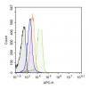
Fig7: Flow cytometric analysis of MMP9 was done on MG-63 cells. The cells were fixed, permeabilized and stained with MMP9 antibody at 1/100 dilution (red) compared with an unlabelled control (cells without incubation with primary antibody; black). After i
特别提示:本公司的所有产品仅可用于科研实验,严禁用于临床医疗及其他非科研用途!

![Anti-MMP9 antibody [10A1]](images/202012/goods_img/92757_G_1607942587940.jpg)