Anti-CAMK4 antibody [B8-C5]
-
概述
- 产品描述The Ca2+/calmodulin-dependent protein kinases (CaM kinases) comprise a structurally related subfamily of serine/threonine kinases which include CaMKI, CaMKII and CaMKIV. CaMKII is a ubiquitously expressed serine/threonine protein kinase that is activated by Ca2+and calmodulin (CaM) and has been implicated in regulation of the cell cycle and transcription. There are four CaMKII isozymes designated α, β, γ and δ, which may or may not be co-expressed in the same tissue type. CaMKIV is stimulated by Ca2+ and CaM but also requires phosphorylation by a CaMK for full activation. Stimulation of the T cell receptor CD3 signaling complex with an anti-CD3 monoclonal antibody leads to a 10-40 fold increase in CaMKIV activity. An additional kinase, CaMKK, functions to activate CaMKI through the specific phosphorylation of the regulatory Threonine residue at position 177.
- 产品名称Anti-CAMK4 antibody [B8-C5]
- 分子量52 kDa
- 种属反应性Human
- 验证应用WB,ICC,IHC-P,FC
- 抗体类型小鼠单抗
- 免疫原Recombinant protein
- 偶联Non-conjugated
-
性能
- 形态Liquid
- 浓度2 mg/mL.
- 存放说明Store at +4℃ after thawing. Aliquot store at -20℃ or -80℃. Avoid repeated freeze / thaw cycles.
- 存储缓冲液1*TBS (pH7.4), 1%BSA, Preservative: 0.05% Sodium Azide.
- 亚型IgG1
- 纯化方式Protein A purified.
- 亚细胞定位Cytoplasm. Nucleus.
- 其它名称
- Brain Ca(2+) calmodulin dependent protein kinase type 4 antibody
- Brain Ca(2+) calmodulin dependent protein kinase type IV antibody
- Brain Ca++-calmodulin dependent protein kinase type IV antibody
more
-
应用
WB: 1:500-1:2,000
ICC: 1:50-1:200
IHC-P: 1:50-1:200
FC: 1:100-1:200
-
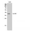
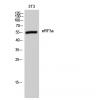
Fig1: Western blot analysis of CAMK4 on human CAMK4 recombinant protein using anti-CAMK4 antibody at 1/1,000 dilution.
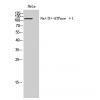
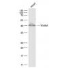
Fig2: Western blot analysis of CAMK4 on HEK293 (1) and CAMK4-hIgGFc transfected HEK293 (2) cell lysate using anti-CAMK4 antibody at 1/1,000 dilution.
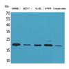
Fig3: Western blot analysis of CAMK4 on different cell lysate using anti-CAMK4 antibody at 1/1,000dilution.
Positive control: Line1:Jurkat Line2:SK-N-SH Line3:Raji Line4:HeLa
Fig4: ICC staining CAMK4 (green) and Actin filaments (red) in HepG2 cells. The nuclear counter stain is DAPI (blue). Cells were fixed in paraformaldehyde, permeabilised with 0.25% Triton X100/PBS.
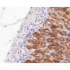
Fig5: Immunohistochemical analysis of paraffin-embedded human ovarian cancer tissue using anti-CAMK4 antibody. Counter stained with hematoxylin.
Fig6: Immunohistochemical analysis of paraffin-embedded human rectum cancer tissue using anti-CAMK4 antibody. Counter stained with hematoxylin.
特别提示:本公司的所有产品仅可用于科研实验,严禁用于临床医疗及其他非科研用途!

![Anti-CAMK4 antibody [B8-C5]](images/202012/goods_img/92771_G_1608032604798.jpg)