Anti-F13A1 antibody [16D1]
-
概述
- 产品描述Hemostasis following tissue injury involves the deployment of essential plasma procoagulants (prothrombin, and factors V, VIII, IX and X), which are involved in a blood coagulation cascade leading to the formation of insoluble fibrin clots and the promotion of platelet aggregation. Coagulation factor VII (serum prothrombin conversion accelerator, proconvertin, F7, Factor VII) is a 406 amino acid, vitamin K-dependent, single chain serine protease that is synthesized in the liver and circulates as an inactive precursor. Factor IXa, factor Xa, factor XIIa, or thrombin mediated proteolytic cleavage of Factor VII at Arg152-Ile153 generates Factor VIIa, an active serine protease composed of a catalytic heavy chain disulfide linked to a light chain, containing two EGF-like domains. Coagulation factor XIII is a terminal effector in the blood coagulation cascade. Plasma factor XIII is a heterotetramer composed of two A subunits and two B subunits. The A subunits have catalytic function, and the noncatalytic B subunits may serve as plasma carrier molecules.
- 产品名称Anti-F13A1 antibody [16D1]
- 分子量83 kDa
-
种属反应性Human
-
验证应用WB,ICC,IHC-P,FC
-
抗体类型小鼠单抗
-
免疫原Recombinant protein within human F13A1 aa 250-500.
-
偶联Non-conjugated
-
性能
-
形态Liquid
-
浓度2 mg/mL.
-
存放说明Store at +4℃ after thawing. Aliquot store at -20℃. Avoid repeated freeze / thaw cycles.
-
存储缓冲液1*PBS (pH7.4), 0.2% BSA, 50% Glycerol. Preservative: 0.05% Sodium Azide.
-
亚型IgG2a
-
纯化方式Protein A affinity purified.
-
亚细胞定位Secreted, cytoplasm.
-
-
其它名称
more
-
bA525O21.1 (coagulation factor XIII, A1 polypeptide) antibody
-
Coagulation factor XIII A chain antibody
-
Coagulation factor XIII A1 polypeptide antibody
-
应用
WB:1:500-1:2,000
ICC:1:50-1:100
IHC-P:1:200-1:1,000
FC:1:50-1:100
-
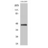
Fig1: Western blot analysis of F13A1 on human placenta tissue lysates. Proteins were transferred to a PVDF membrane and blocked with 5% BSA in PBS for 1 hour at room temperature. The primary antibody was used in 5% BSA at room temperature for 2 hours. Goat Anti-Mouse IgG - HRP Secondary Antibody (HA1006) at 1:5,000 dilution was used for 1 hour at room temperature.
Fig2: ICC staining of F13A1 in MCF-7 cells (green). Formalin fixed cells were permeabilized with 0.1% Triton X-100 in TBS for 10 minutes at room temperature and blocked with 1% Blocker BSA for 15 minutes at room temperature. Cells were probed with the primary antibody for 1 hour at room temperature, washed with PBS. Alexa Fluor®488 Goat anti-Mouse IgG was used as the secondary antibody at 1/1,000 dilution. The nuclear counter stain is DAPI (blue).
Fig3: ICC staining of F13A1 in MG-63 cells (green). Formalin fixed cells were permeabilized with 0.1% Triton X-100 in TBS for 10 minutes at room temperature and blocked with 1% Blocker BSA for 15 minutes at room temperature. Cells were probed with the primary antibodyfor 1 hour at room temperature, washed with PBS. Alexa Fluor®488 Goat anti-Mouse IgG was used as the secondary antibody at 1/1,000 dilution. The nuclear counter stain is DAPI (blue).
Fig4: Immunohistochemical analysis of paraffin-embedded human placenta tissue using anti-F13A1 antibody. The section was pre-treated using heat mediated antigen retrieval with Tris-EDTA buffer (pH 8.0-8.4) for 20 minutes.The tissues were blocked in 5% BSA for 30 minutes at room temperature, washed with ddH2O and PBS, and then probed with the primary antibody for 30 minutes at room temperature. The detection was performed using an HRP conjugated compact polymer system. DAB was used as the chromogen. Tissues were counterstained with hematoxylin and mounted with DPX.
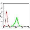
Fig5: Flow cytometric analysis of F13A1 was done on A549 cells. The cells were fixed, permeabilized and stained with the primary antibody (red). After incubation of the primary antibody at room temperature for an hour, the cells were stained with a Alexa Fluor 488-conjugated Goat anti-Mouse IgG Secondary antibody at 1/1000 dilution for 30 minutes.Unlabelled sample was used as a control (cells without incubation with primary antibody; black).
特别提示:本公司的所有产品仅可用于科研实验,严禁用于临床医疗及其他非科研用途!

![Anti-F13A1 antibody [16D1]](images/202012/goods_img/92823_G_1608035421674.jpg)