Anti-PLD2 antibody [D9-E4]
-
概述
- 产品描述The protein encoded by this gene catalyzes the hydrolysis of phosphatidylcholine to phosphatidic acid and choline. The activity of the encoded enzyme is enhanced by phosphatidylinositol 4,5-bisphosphate and ADP-ribosylation factor-1. This protein localizes to the peripheral membrane and may be involved in cytoskeletal organization, cell cycle control, transcriptional regulation, and/or regulated secretion. Two transcript variants encoding different isoforms have been found for this gene.
- 产品名称Anti-PLD2 antibody [D9-E4]
- 分子量106 kDa
- 种属反应性Human
- 验证应用WB,ICC,IHC-P,FC
- 抗体类型小鼠单抗
- 免疫原Recombinant protein
- 偶联Non-conjugated
-
性能
- 形态Liquid
- 浓度2 mg/mL.
- 存放说明Store at +4℃ after thawing. Aliquot store at -20℃ or -80℃. Avoid repeated freeze / thaw cycles.
- 存储缓冲液1*TBS (pH7.4), 1%BSA, 40%Glycerol. Preservative: 0.05% Sodium Azide.
- 亚型IgG1
- 纯化方式Protein A purified.
- 亚细胞定位Membrane. Peripheral membrane protein.
- 其它名称
- Choline phosphatase 2 antibody
- EC 3.1.4.4 antibody
- hPLD 2 antibody
more
-
应用
WB: 1:500-1:2,000
ICC: 50-1:200
IHC-P: 1:50-1:200
FC: 1:50-1:200
-
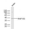
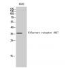
Fig1: Western blot analysis of PLD2 on human PLD2 recombinant protein using anti-PLD2 antibody at 1/1,000 dilution.
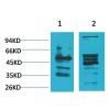
Fig2: Western blot analysis of PLD2 on HEK293 (1) and PLD2-hIgGFc transfected HEK293 (2) cell lysate using anti-PLD2 antibody at 1/1,000 dilution.
Fig3: ICC staining PLD2 (green) and Actin filaments (red) in MCF-7 cells. The nuclear counter stain is DAPI (blue). Cells were fixed in paraformaldehyde, permeabilised with 0.25% Triton X100/PBS.
Fig4: ICC staining PLD2 (green) and Actin filaments (red) in SK-OV-3 cells. The nuclear counter stain is DAPI (blue). Cells were fixed in paraformaldehyde, permeabilised with 0.25% Triton X100/PBS.
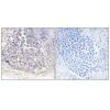
Fig5: Immunohistochemical analysis of paraffin-embedded human renal tissues using anti-PLD2 antibody. Counter stained with hematoxylin.
Fig6: Immunohistochemical analysis of paraffin-embedded human esophageal cancer tissues using anti-PLD2 antibody. Counter stained with hematoxylin.
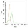
Fig7: Flow cytometric analysis of Hela cells with PLD2 antibody at 1/100 dilution (green) compared with an unlabelled control (cells without incubation with primary antibody; red).
特别提示:本公司的所有产品仅可用于科研实验,严禁用于临床医疗及其他非科研用途!

![Anti-PLD2 antibody [D9-E4]](images/202012/goods_img/92839_G_1608036272634.jpg)