Anti-HLA-DR antibody [10-D8]
-
概述
- 产品描述Major histocompatibility complex (MHC) class II molecules destined for presentation to CD4+ helper T cells is determined by two key events. These events include the dissociation of class II-associated invariant chain peptides (CLIP) from an antigen binding groove in mhc ii-a/b dimers through the activity of MHC molecules HLA-DM and -DO, and subsequent peptide antigen binding. Accumulating in endosomal/lysosomal compartments and on the surface of B cells, HLA-DM, -DO molecules regulate the dissociation of CLIP and the subsequent binding of exogenous peptides to HLA class II molecules (HLA-DR, -DQ and -DP) by sustaining a conformation that favors peptide exchange. RFLP analysis of HLA-DM genes from rheumatoid arthritis (RA) patients suggests that certain polymorphisms are genetic factors for RA susceptibility. HLA-B belongs to the HLA class I heavy chain paralogs. Class I molecules play a central role in the immune system by presenting peptides derived from the endoplasmic reticulum lumen. HLA-B and -C can form heterodimers consisting of a membrane anchored heavy chain and a light chain (β-2-Microglobulin). Polymorphisms yield hundreds of HLA-B and -C alleles.
- 产品名称Anti-HLA-DR antibody [10-D8]
- 分子量29 kDa
- 种属反应性Human
- 验证应用WB,IHC-P,FC
- 抗体类型小鼠单抗
- 免疫原Peptide
- 偶联Non-conjugated
-
性能
- 形态Liquid
- 浓度2 mg/mL.
- 存放说明Store at +4℃ after thawing. Aliquot store at -20℃ or -80℃. Avoid repeated freeze / thaw cycles.
- 存储缓冲液1*PBS (pH7.4), 0.2% BSA, 50% Glycerol. Preservative: 0.05% Sodium Azide.
- 亚型IgG1
- 纯化方式Peptide affinity purified
- 亚细胞定位Cell membrane. Endoplasmic reticulum membrane.
- 其它名称
- DR alpha chain antibody
- DR alpha chain precursor antibody
- DRA_HUMAN antibody
more
-
应用
WB: 1:500-1:1,000
IHC-P: 1:50-1:200
FC: 1:50-1:200
-
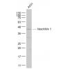
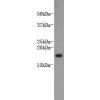
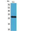
Fig1: Western blot analysis of HLA-DR on Raji cell lysate using anti-HLA-DR antibody at 1/1,000 dilution.
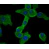
Fig2: Immunohistochemical analysis of paraffin-embedded human tonsil tissue using anti-HLA-DR antibody. Counter stained with hematoxylin.
Fig3: Immunohistochemical analysis of paraffin-embedded human lung cancer tissue using anti-HLA-DR antibody. Counter stained with hematoxylin.
Fig4: Immunohistochemical analysis of paraffin-embedded human colon cancer tissue using anti-HLA-DR antibody. Counter stained with hematoxylin.
Fig5: Immunohistochemical analysis of paraffin-embedded human skin tissue using anti-HLA-DR antibody. Counter stained with hematoxylin.
Fig6: Immunohistochemical analysis of paraffin-embedded human kidney tissue using anti-HLA-DR antibody. Counter stained with hematoxylin.
Fig7: Flow cytometric analysis of Daudi cells with HLA-DR antibody at 1/100 dilution (red) compared with an unlabelled control (cells without incubation with primary antibody; black).
特别提示:本公司的所有产品仅可用于科研实验,严禁用于临床医疗及其他非科研用途!

![Anti-HLA-DR antibody [10-D8]](images/202012/goods_img/92933_G_1608203458851.jpg)