Anti-MTA2 antibody [17A2]
-
概述
- 产品描述This gene encodes a protein that has been identified as a component of NuRD, a nucleosome remodeling deacetylase complex identified in the nucleus of human cells. It shows a very broad expression pattern and is strongly expressed in many tissues. It may represent one member of a small gene family that encode different but related proteins involved either directly or indirectly in transcriptional regulation. Their indirect effects on transcriptional regulation may include chromatin remodeling. It is closely related to another member of this family, a protein that has been correlated with the metastatic potential of certain carcinomas. These two proteins are so closely related that they share the same types of domains. These domains include two DNA binding domains, a dimerization domain, and a domain commonly found in proteins that methylate DNA. One of the proteins known to be a target protein for this gene product is p53. Deacetylation of p53 is correlated with a loss of growth inhibition in transformed cells supporting a connection between these gene family members and metastasis.
- 产品名称Anti-MTA2 antibody [17A2]
- 分子量75 kDa
- 种属反应性Human
- 验证应用WB,IHC-P,FC
- 抗体类型小鼠单抗
- 免疫原Recombinant protein within human MTA2 aa 50-300.
- 偶联Non-conjugated
-
性能
- 形态Liquid
- 浓度2 mg/mL.
- 存放说明Store at +4℃ after thawing. Aliquot store at -20℃. Avoid repeated freeze / thaw cycles.
- 存储缓冲液1*PBS (pH7.4), 0.2% BSA, 50% Glycerol. Preservative: 0.05% Sodium Azide.
- 亚型IgG2b
- 纯化方式Protein G purified.
- 亚细胞定位Nucleus.
- 其它名称
- DKFZp686F2281 antibody
- Mata1l1 antibody
- Metastasis associated 1 family member 2 antibody
more
-
应用
WB:1:500-1:2,000
IHC-P:1:500-1:1,000
FC:1:50-1:100
-
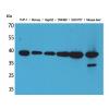
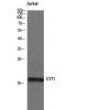
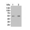
Fig1: Western blot analysis of MTA2 on different lysates. Proteins were transferred to a PVDF membrane and blocked with 5% BSA in PBS for 1 hour at room temperature. The primary antibody (was used in 5% BSA at room temperature for 2 hours. Goat Anti-Mouse IgG - HRP Secondary Antibody (HA1006) at 1:5,000 dilution was used for 1 hour at room temperature.
Positive control:
Lane 1: K562 cell lysate
Lane 2: MCF-7 cell lysate
Lane 3: SH-SY5Y cell lysate
Lane 4: Daudi cell lysate
Fig2: Immunohistochemical analysis of paraffin-embedded human tonsil tissue using anti-MTA2 antibody. The section was pre-treated using heat mediated antigen retrieval with sodium citrate buffer (pH 6.0) for 20 minutes. The tissues were blocked in 5% BSA for 30 minutes at room temperature, washed with ddH2O and PBS, and then probed with the primary antibody ( for 30 minutes at room temperature. The detection was performed using an HRP conjugated compact polymer system. DAB was used as the chromogen. Tissues were counterstained with hematoxylin and mounted with DPX.
Fig3: Immunohistochemical analysis of paraffin-embedded human colon carcinoma tissue using anti-MTA2 antibody. The section was pre-treated using heat mediated antigen retrieval with sodium citrate buffer (pH 6.0) for 20 minutes. The tissues were blocked in 5% BSA for 30 minutes at room temperature, washed with ddH2O and PBS, and then probed with the primary antibody for 30 minutes at room temperature. The detection was performed using an HRP conjugated compact polymer system. DAB was used as the chromogen. Tissues were counterstained with hematoxylin and mounted with DPX.
Fig4: Immunohistochemical analysis of paraffin-embedded human skin tissue using anti-MTA2 antibody. The section was pre-treated using heat mediated antigen retrieval with sodium citrate buffer (pH 6.0) for 20 minutes. The tissues were blocked in 5% BSA for 30 minutes at room temperature, washed with ddH2O and PBS, and then probed with the primary antibody for 30 minutes at room temperature. The detection was performed using an HRP conjugated compact polymer system. DAB was used as the chromogen. Tissues were counterstained with hematoxylin and mounted with DPX.
Fig5: Immunohistochemical analysis of paraffin-embedded human breast tissue using anti-MTA2 antibody. The section was pre-treated using heat mediated antigen retrieval with sodium citrate buffer (pH 6.0) for 20 minutes. The tissues were blocked in 5% BSA for 30 minutes at room temperature, washed with ddH2O and PBS, and then probed with the primary antibody (for 30 minutes at room temperature. The detection was performed using an HRP conjugated compact polymer system. DAB was used as the chromogen. Tissues were counterstained with hematoxylin and mounted with DPX.
Fig6: Immunohistochemical analysis of paraffin-embedded human breast carcinoma tissue using anti-MTA2 antibody. The section was pre-treated using heat mediated antigen retrieval with sodium citrate buffer (pH 6.0) for 20 minutes. The tissues were blocked in 5% BSA for 30 minutes at room temperature, washed with ddH2O and PBS, and then probed with the primary antibody for 30 minutes at room temperature. The detection was performed using an HRP conjugated compact polymer system. DAB was used as the chromogen. Tissues were counterstained with hematoxylin and mounted with DPX.
Fig7: Immunohistochemical analysis of paraffin-embedded human esophagus tissue using anti-MTA2 antibody. The section was pre-treated using heat mediated antigen retrieval with sodium citrate buffer (pH 6.0) for 20 minutes. The tissues were blocked in 5% BSA for 30 minutes at room temperature, washed with ddH2O and PBS, and then probed with the primary antibody (for 30 minutes at room temperature. The detection was performed using an HRP conjugated compact polymer system. DAB was used as the chromogen. Tissues were counterstained with hematoxylin and mounted with DPX.
Fig8: Immunohistochemical analysis of paraffin-embedded human pancreas tissue using anti-MTA2 antibody. The section was pre-treated using heat mediated antigen retrieval with sodium citrate buffer (pH 6.0) for 20 minutes. The tissues were blocked in 5% BSA for 30 minutes at room temperature, washed with ddH2O and PBS, and then probed with the primary antibody ( for 30 minutes at room temperature. The detection was performed using an HRP conjugated compact polymer system. DAB was used as the chromogen. Tissues were counterstained with hematoxylin and mounted with DPX.
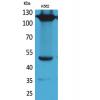
Fig9: Flow cytometric analysis of MTA2 was done on MCF-7 cells. The cells were fixed, permeabilized and stained with the primary antibody (red). After incubation of the primary antibody at room temperature for an hour, the cells were stained with a Alexa Fluor 488-conjugated Goat anti-Mouse IgG Secondary antibody at 1/1000 dilution for 30 minutes.Unlabelled sample was used as a control (cells without incubation with primary antibody; black).
特别提示:本公司的所有产品仅可用于科研实验,严禁用于临床医疗及其他非科研用途!

![Anti-MTA2 antibody [17A2]](images/202012/goods_img/92944_G_1608204330307.jpg)