Anti-CD166 antibody [5-5]
-
概述
- 产品描述Cell adhesion molecule that mediates both heterotypic cell-cell contacts via its interaction with CD6, as well as homotypic cell-cell contacts . Promotes T-cell activation and proliferation via its interactions with CD6 . Contributes to the formation and maturation of the immunological synapse via its interactions with CD6 . Mediates homotypic interactions with cells that express ALCAM . Required for normal hematopoietic stem cell engraftment in the bone marrow . Mediates attachment of dendritic cells onto endothelial cells via homotypic interaction . Inhibits endothelial cell migration and promotes endothelial tube formation via homotypic interactions . Required for normal organization of the lymph vessel network. Required for normal hematopoietic stem cell engraftment in the bone marrow. Plays a role in hematopoiesis; required for normal numbers of hematopoietic stem cells in bone marrow. Promotes in vitro osteoblast proliferation and differentiation (By similarity). Promotes neurite extension, axon growth and axon guidance; axons grow preferentially on surfaces that contain ALCAM. Mediates outgrowth and pathfinding for retinal ganglion cell axons (By similarity).
- 产品名称Anti-CD166 antibody [5-5]
- 分子量Predicted band size: 65 kDa.
- 种属反应性Human
- 验证应用WB,IHC-P,ICC,FC
- 抗体类型小鼠单抗
- 免疫原Recombinant protein within human CD166 aa 1-300.
- 偶联Non-conjugated
-
性能
- 形态Liquid
- 浓度2 mg/ml.
- 存放说明Store at +4℃ after thawing. Aliquot store at -20℃. Avoid repeated freeze / thaw cycles.
- 存储缓冲液1*TBS (pH7.4), 1%BSA, 50%Glycerol. Preservative: 0.05% Sodium Azide.
- 亚型IgG1
- 纯化方式Protein A purified.
- 亚细胞定位Cell membrane, Cell projection, Membrane, Secreted
- 其它名称
- Activated leukocyte cell adhesion molecule antibody
- ALCAM antibody
- ALCAM protein antibody
more
-
应用
WB: 1:500-1:1,000
IHC-P: 1:50-1:200
ICC: 1:50-1:100
FC: 1:50-1:100
-
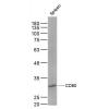
Fig1: Western blot analysis of CD166 on different lysates. Proteins were transferred to a PVDF membrane and blocked with 5% BSA in PBS for 1 hour at room temperature. The primary antibody ( was used in 5% BSA at room temperature for 2 hours. Goat Anti-Mouse IgG - HRP Secondary Antibody (HA1006) at 1:5,000 dilution was used for 1 hour at room temperature.
Positive control:
Lane 1: PC-3M cell lysate
Lane 2: A549 cell lysate
Lane 3: SHSY5Y cell lysate
Fig2: ICC staining of CD166 in A431 cells (green). Formalin fixed cells were permeabilized with 0.1% Triton X-100 in TBS for 10 minutes at room temperature and blocked with 1% Blocker BSA for 15 minutes at room temperature. Cells were probed with the primary antibody for 1 hour at room temperature, washed with PBS. Alexa Fluor®488 Goat anti-Rabbit IgG was used as the secondary antibody at 1/100 dilution. The nuclear counter stain is DAPI (blue).
Fig3: ICC staining of CD166 in Huvec cells (green). Formalin fixed cells were permeabilized with 0.1% Triton X-100 in TBS for 10 minutes at room temperature and blocked with 1% Blocker BSA for 15 minutes at room temperature. Cells were probed with the primary antibody for 1 hour at room temperature, washed with PBS. Alexa Fluor®488 Goat anti-Mouse IgG was used as the secondary antibody at 1/100 dilution. The nuclear counter stain is DAPI (blue).
Fig4: Immunohistochemical analysis of paraffin-embedded human liver tissue using anti-CD166 antibody. The section was pre-treated using heat mediated antigen retrieval with Tris-EDTA buffer (pH 8.0-8.4) for 20 minutes.The tissues were blocked in 5% BSA for 30 minutes at room temperature, washed with ddH2O and PBS, and then probed with the primary antibody for 30 minutes at room temperature. The detection was performed using an HRP conjugated compact polymer system. DAB was used as the chromogen. Tissues were counterstained with hematoxylin and mounted with DPX.
Fig5: Flow cytometric analysis of CD166 was done on THP-1 cells. The cells were fixed, permeabilized and stained with the primary antibody(red). After incubation of the primary antibody at room temperature for an hour, the cells were stained with a Alexa Fluor 488-conjugated goat anti-Mouse IgG Secondary antibody at 1/500 dilution for 30 minutes.Unlabelled sample was used as a control (cells without incubation with primary antibody; black).
特别提示:本公司的所有产品仅可用于科研实验,严禁用于临床医疗及其他非科研用途!

![Anti-CD166 antibody [5-5]](images/202012/goods_img/92950_G_1608204793725.jpg)