Anti-CD68 antibody [A3C4]
-
概述
- 产品描述CD68 (Cluster of Differentiation 68) is a protein highly expressed by cells in the monocyte lineage (e.g., monocytic phagocytes, osteoclasts), by circulating macrophages, and by tissue macrophages (e.g., Kupffer cells, microglia). Human CD68 is a transmembrane glycoprotein, heavily glycosylated in its extracellular domain, with a molecular weight of 110 kD. Its primary sequence consists of 354 amino acids with predicted molecular weight of 37.4 kD if it were not glycosylated. Immunohistochemistry can be used to identify the presence of CD68, which is found in the cytoplasmic granules of a range of different blood cells and myocytes. It is particularly useful as a marker for the various cells of the macrophage lineage, including monocytes, histiocytes, giant cells, Kupffer cells, and osteoclasts. This allows it to be used to distinguish diseases of otherwise similar appearance, such as the monocyte/macrophage and lymphoid forms of leukaemia (the latter being CD68 negative). Its presence in macrophages also makes it useful in diagnosing conditions related to proliferation or abnormality of these cells, such as malignant histiocytosis, histiocytic lymphoma, and Gaucher's disease. Anti-CD68 monoclonal antibodies that react with tissues of rodent and other species include ED1, FA-11, KP1 (a.k.a. C68/684), 6A326, 6F3, 12E2, 10B1909, and SPM130. Monoclonals that react with humans include, Ki-M7, PG-M1, 514H12, ABM53F5, 3F7C6, 3F7D3, Y1/82A, EPR20545, CDLA68-1, LAMP4-824.
- 产品名称Anti-CD68 antibody [A3C4]
- 分子量Predicted band size: 37 kDa.
- 种属反应性Human
- 验证应用WB,IHC-P,FC
- 抗体类型小鼠单抗
- 免疫原Synthetic peptide within human CD68 aa 320-354.
- 偶联Non-conjugated
-
性能
- 形态Liquid
- 浓度2 mg/mL.
- 存放说明Store at +4℃ after thawing. Aliquot store at -20℃. Avoid repeated freeze / thaw cycles.
- 存储缓冲液1*PBS (pH7.4), 0.2% BSA, 50% Glycerol. Preservative: 0.05% Sodium Azide.
- 亚型IgG1
- 纯化方式Protein G affinity purified.
- 亚细胞定位Cell membrane. Endosome membrane, lysosome membrane.
- 其它名称
- CD 68 antibody
- CD68 antibody
- CD68 antigen antibody
more
-
应用
WB:1:1,000-1:2,000
IHC-P:1:50-1:200
FC:1:50-1:100
-
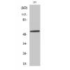
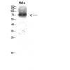
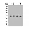
Fig1: Western blot analysis of CD68 on different lysates. Proteins were transferred to a PVDF membrane and blocked with 5% BSA in PBS for 1 hour at room temperature. The primary antibody ( was used in 5% BSA at room temperature for 2 hours. Goat Anti-Mouse IgG - HRP Secondary Antibody (HA1001) at 1:5,000 dilution was used for 1 hour at room temperature.
Positive control:
Lane 1: A431 cell lysate
Lane 2: THP-1 cell lysate
Fig2: Immunohistochemical analysis of paraffin-embedded human tonsil tissue using anti-CD68 antibody. The section was pre-treated using heat mediated antigen retrieval with Tris-EDTA buffer (pH 8.0-8.4) for 20 minutes.The tissues were blocked in 5% BSA for 30 minutes at room temperature, washed with ddH2O and PBS, and then probed with the primary antibody ( for 30 minutes at room temperature. The detection was performed using an HRP conjugated compact polymer system. DAB was used as the chromogen. Tissues were counterstained with hematoxylin and mounted with DPX.
Fig3: Immunohistochemical analysis of paraffin-embedded human spleen tissue using anti-CD68 antibody. The section was pre-treated using heat mediated antigen retrieval with Tris-EDTA buffer (pH 8.0-8.4) for 20 minutes.The tissues were blocked in 5% BSA for 30 minutes at room temperature, washed with ddH2O and PBS, and then probed with the primary antibody for 30 minutes at room temperature. The detection was performed using an HRP conjugated compact polymer system. DAB was used as the chromogen. Tissues were counterstained with hematoxylin and mounted with DPX.
Fig4: Immunohistochemical analysis of paraffin-embedded human lung tissue using anti-CD68 antibody. The section was pre-treated using heat mediated antigen retrieval with Tris-EDTA buffer (pH 8.0-8.4) for 20 minutes.The tissues were blocked in 5% BSA for 30 minutes at room temperature, washed with ddH2O and PBS, and then probed with the primary antibody (for 30 minutes at room temperature. The detection was performed using an HRP conjugated compact polymer system. DAB was used as the chromogen. Tissues were counterstained with hematoxylin and mounted with DPX.
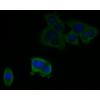
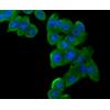
Fig5: Flow cytometric analysis of CD68 was done on THP-1 cells. The cells were fixed, permeabilized and stained with the primary antibody(red). After incubation of the primary antibody at room temperature for an hour, the cells were stained with a Alexa Fluor 488-conjugated Goat anti-Mouse IgG Secondary antibody at 1/1000 dilution for 30 minutes.Unlabelled sample was used as a control (cells without incubation with primary antibody; black).
特别提示:本公司的所有产品仅可用于科研实验,严禁用于临床医疗及其他非科研用途!

![Anti-CD68 antibody [A3C4]](images/202012/goods_img/92957_G_1608462456613.jpg)