Anti-CD163 antibody [A3B3]
-
概述
- 产品描述Acute phase-regulated receptor involved in clearance and endocytosis of hemoglobin/haptoglobin complexes by macrophages and may thereby protect tissues from free hemoglobin-mediated oxidative damage. Exhibits a higher affinity for complexes of hemoglobin and multimeric haptoglobin of HP*1F phenotype than for complexes of hemoglobin and dimeric haptoglobin of HP*1S phenotype. Induces a cascade of intracellular signals that involves tyrosine kinase-dependent calcium mobilization, inositol triphosphate production and secretion of IL6 and CSF1. After shedding, the soluble form (sCD163) may play an anti-inflammatory role, and may be a valuable diagnostic parameter for monitoring macrophage activation in inflammatory conditions. Intravenous lipopolysaccharide (LPS) produces a rapid rise of sCD163 in plasma of patient as it induces metalloproteinase-mediated shedding from monocytes surface. The soluble form (sCD163) in plasma is a novel parameter in diseases affecting macrophage function and monocyte/macrophage load in the body. The concentration of sCD163 is probably reflecting the number of macrophages of the 'alternative macrophage activation' phenotype with a high CD163 expression playing a major role in dampening the inflammatory response and scavenging components of damaged cells. This has initiated a number of clinical studies for evaluation of sCD163 as a disease marker in inflammatory conditions e.g. infection, autoimmune disease, transplantation, atherosclerosis and cancer.
- 产品名称Anti-CD163 antibody [A3B3]
- 分子量Predicted band size: 125 kDa.
- 种属反应性Human
- 验证应用WB,IHC-P,FC
- 抗体类型小鼠单抗
- 免疫原Recombinant protein within human CD163 aa 1-170.
- 偶联Non-conjugated
-
性能
- 形态Liquid
- 浓度2 mg/mL.
- 存放说明Store at +4℃ after thawing. Aliquot store at -20℃. Avoid repeated freeze / thaw cycles.
- 存储缓冲液1*PBS (pH7.4), 0.2% BSA, 50% Glycerol. Preservative: 0.05% Sodium Azide.
- 亚型IgG1
- 纯化方式Protein G affinity purified.
- 亚细胞定位Secreted, cell membrane.
- 其它名称
- C163A_HUMAN antibody
- CD 163 antibody
- CD163 antibody
more
-
应用
WB:1:500-1:2,000
IHC-P:1:100-1:500
FC:1:50-1:100
-
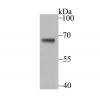
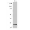
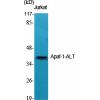
Fig1: Western blot analysis of CD163 on human liver tissue lysate (1) and human thymus tissue lysate (2). Proteins were transferred to a PVDF membrane and blocked with 5% BSA in PBS for 1 hour at room temperature. The primary antibody (was used in 5% BSA at room temperature for 2 hours. Goat Anti-Mouse IgG - HRP Secondary Antibody (HA1001) at 1:5,000 dilution was used for 1 hour at room temperature.
Fig2: Immunohistochemical analysis of paraffin-embedded human lung tissue using anti-CD163 antibody. The section was pre-treated using heat mediated antigen retrieval with Tris-EDTA buffer (pH 8.0-8.4) for 20 minutes.The tissues were blocked in 5% BSA for 30 minutes at room temperature, washed with ddH2O and PBS, and then probed with the primary antibody for 30 minutes at room temperature. The detection was performed using an HRP conjugated compact polymer system. DAB was used as the chromogen. Tissues were counterstained with hematoxylin and mounted with DPX.
Fig3: Immunohistochemical analysis of paraffin-embedded human liver carcinoma tissue using anti-CD163 antibody. The section was pre-treated using heat mediated antigen retrieval with Tris-EDTA buffer (pH 8.0-8.4) for 20 minutes.The tissues were blocked in 5% BSA for 30 minutes at room temperature, washed with ddH2O and PBS, and then probed with the primary antibody for 30 minutes at room temperature. The detection was performed using an HRP conjugated compact polymer system. DAB was used as the chromogen. Tissues were counterstained with hematoxylin and mounted with DPX.
Fig4: Immunohistochemical analysis of paraffin-embedded human colom tissue using anti-CD163 antibody. The section was pre-treated using heat mediated antigen retrieval with Tris-EDTA buffer (pH 8.0-8.4) for 20 minutes.The tissues were blocked in 5% BSA for 30 minutes at room temperature, washed with ddH2O and PBS, and then probed with the primary antibody for 30 minutes at room temperature. The detection was performed using an HRP conjugated compact polymer system. DAB was used as the chromogen. Tissues were counterstained with hematoxylin and mounted with DPX.
Fig5: Immunohistochemical analysis of paraffin-embedded human placenta tissue using anti-CD163 antibody. The section was pre-treated using heat mediated antigen retrieval with Tris-EDTA buffer (pH 8.0-8.4) for 20 minutes.The tissues were blocked in 5% BSA for 30 minutes at room temperature, washed with ddH2O and PBS, and then probed with the primary antibody for 30 minutes at room temperature. The detection was performed using an HRP conjugated compact polymer system. DAB was used as the chromogen. Tissues were counterstained with hematoxylin and mounted with DPX.
Fig6: Flow cytometric analysis of CD163 was done on HT-29 cells. The cells were fixed, permeabilized and stained with the primary antibody (red). After incubation of the primary antibody at room temperature for an hour, the cells were stained with a Alexa Fluor 488-conjugated Goat anti-Mouse IgG Secondary antibody at 1/1000 dilution for 30 minutes.Unlabelled sample was used as a control (cells without incubation with primary antibody; black).
特别提示:本公司的所有产品仅可用于科研实验,严禁用于临床医疗及其他非科研用途!

![Anti-CD163 antibody [A3B3]](images/202012/goods_img/92961_G_1608462694861.jpg)