Anti-Vang-like protein 1 antibody
-
概述
- 产品描述Vang-like protein 1 is a multi-pass membrane protein, expressed specifically in testis and ovary. It interacts through its C-terminal region with the N-terminal half of DVL1, DVL2 and DVL3. Vangl proteins play a key developmental role in establishing planar cell polarity (PCP) and in regulating convergent extension (CE) movements during embryogenesis. Vangl1 and Vangl2 are both downregulated in several cancer cell lines and primary tumors. Defects in VANGL1 are a cause of neural tube defects (NTD) and a cause of sacral defect with anterior meningocele (SDAM).
- 产品名称Anti-Vang-like protein 1 antibody
- 分子量80 kDa
- 种属反应性Human
- 验证应用WB,ICC
- 抗体类型兔多抗
- 免疫原Synthetic peptide within human 100-160 aa.
- 偶联Non-conjugated
-
性能
- 形态Liquid
- 浓度1 mg/mL.
- 存放说明Store at +4℃ after thawing. Aliquot store at -20℃. Avoid repeated freeze / thaw cycles.
- 存储缓冲液1*PBS (pH7.4), 0.2% BSA, 25% Glycerol. Preservative: 0.05% Sodium Azide.
- 亚型IgG
- 纯化方式Peptide affinity purified.
- 亚细胞定位Cell membrane. Membrane.
- 其它名称ANGL planar cell polarity protein 1 antibody
KAI1 C-terminal interacting tetraspanin antibody
KITENIN antibody
Loop tail protein 2 homolog antibody
Loop-tail protein 2 homolog antibody
LPP2 antibody
MGC5338 antibody
STB2 antibody
STBM2 antibody
Strabismus 2 antibody
Strabismus, Drosophila, homolog of, 2 antibody
Van Gogh like protein 1 antibody
Van Gogh, Drosophila, homolog of, 1 antibody
Van Gogh-like protein 1 antibody
Vang like 1 (van gogh, Drosophila) antibody
Vang like protein 1 antibody
Vang-like 1 antibody
Vang-like protein 1 antibody
VANG1_HUMAN antibody
VANGL 1 antibody
Vangl1 antibody
more
-
应用
WB: 1:500
ICC: 1:100-1:200
-
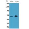
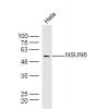
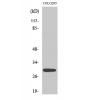
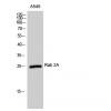
Fig1: Western blot analysis of VANGL1 on MCF-7 cell lysate using anti-VANGL1 antibody at 1/500 dilution.
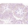
Fig2: ICC staining VANGL1 in Hela cells (green). The nuclear counter stain is DAPI (blue). Cells were fixed in paraformaldehyde, permeabilised with 0.25% Triton X100/PBS.
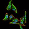
Fig3: ICC staining VANGL1 in LOVO cells (green). The nuclear counter stain is DAPI (blue). Cells were fixed in paraformaldehyde, permeabilised with 0.25% Triton X100/PBS.
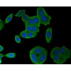
Fig4:
ICC staining VANGL1in MCF-7 cells (green). The nuclear counter stain is DAPI (blue). Cells were fixed in paraformaldehyde, permeabilised with 0.25% Triton X100/PBS.
特别提示:本公司的所有产品仅可用于科研实验,严禁用于临床医疗及其他非科研用途!