Anti-BAX antibody
-
概述
- 产品描述BAX is a member of the Bcl-2 gene family. Apoptosis regulator BAX promotes apoptosis by binding to and antagonizing the Bcl-2 protein. In healthy mammalian cells, the majority of BAX is found in the cytosol, but upon initiation of apoptotic signaling, Bax undergoes a conformational shift. Upon induction of apoptosis, BAX becomes organelle membrane-associated, and in particular, mitochondrial membrane associated. The expression of BAX is upregulated by the tumor suppressor protein p53, and BAX has been shown to be involved in p53-mediated apoptosis. The p53 protein is a transcription factor that, when activated as part of the cell's response to stress, regulates many downstream target genes, including BAX.
- 产品名称Anti-BAX antibody
- 分子量21 kDa
- 种属反应性Human,Mouse,Rat
- 验证应用WB,ICC,IHC-P
- 抗体类型兔多抗
- 免疫原This antibody is produced by immunizing rabbits with a synthetic peptide (KLH-coupled) corresponding to N-terminal BAX.
- 偶联Non-conjugated
-
性能
- 形态Liquid
- 浓度1 mg/mL.
- 存放说明Store at +4℃ after thawing. Aliquot store at -20℃ or -80℃. Avoid repeated freeze / thaw cycles.
- 存储缓冲液1*PBS (pH7.4), 0.2% BSA, 40% Glycerol. Preservative: 0.05% Sodium Azide.
- 亚型IgG
- 纯化方式Peptide affinity purified
- 亚细胞定位Mitochondrion membrane, cytoplasm
- 其它名称Apoptosis regulator BAX antibody
BAX antibody
Bax-protein antibody
BAX_HUMAN antibody
BAXA antibody
Baxdelta2G9 antibody
Baxdelta2G9omega antibody
Baxdelta2omega antibody
Bcl-2-like protein 4 antibody
BCL2 associated X protein antibody
BCL2 associated X protein omega antibody
BCL2 associated X protein transcript variant delta2 antibody
Bcl2-L-4 antibody
BCL2L4 antibody
membrane isoform alpha antibody
more
-
应用
WB: 1:500-1:1,000
ICC: 1:200
IHC-P: 1:200
-
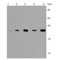
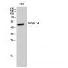
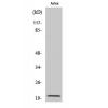
Fig1: Western blot analysis of Bax on different lysates using anti-Bax antibody at 1/500 dilution.
Positive control:
Lane 1: HepG2 Lane2 :Hela Lane 3:Mouse embryonic stem cell Lane 4:PC12 Lane 5:F9
Fig2: ICC staining Bax in HepG2 cells (green). The nuclear counter stain is DAPI (blue).Cells were fixed in paraformaldehyde, permeabilised with 0.25% Triton X100/PBS.
Fig3: ICC staining Bax in 293 cells (red). The nuclear counter stain is DAPI (blue). Cells were fixed in paraformaldehyde, permeabilised with 0.25% Triton X100/PBS.
Fig4: ICC staining Bax in F9 cells (green). The nuclear counter stain is DAPI (blue). Cells were fixed in paraformaldehyde, permeabilised with 0.25% Triton X100/PBS.
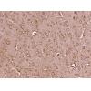
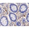
Fig5: Immunohistochemical analysis of paraffin-embedded human gall bladder tissue using anti-Bax antibody. Counter stained with hematoxylin.
Fig6: Immunohistochemical analysis of paraffin-embedded human cervical tissue using anti-Bax antibody. Counter stained with hematoxylin.
Fig7: Immunohistochemical analysis of paraffin-embedded mouse testis tissue using anti-Bax antibody. Counter stained with hematoxylin.
特别提示:本公司的所有产品仅可用于科研实验,严禁用于临床医疗及其他非科研用途!