Anti-Nfic antibody
-
概述
- 产品描述Recognizes and binds the palindromic sequence 5'-TTGGCNNNNNGCCAA-3' present in viral and cellular promoters and in the origin of replication of adenovirus type 2. These proteins are individually capable of activating transcription and replication.
- 产品名称Anti-Nfic antibody
- 分子量55 kDa
- 种属反应性Human
- 验证应用WB,ICC,IHC-P,FC
- 抗体类型兔多抗
- 免疫原Synthetic peptide within human Nfic 400-450 aa.
- 偶联Non-conjugated
-
性能
- 形态Liquid
- 浓度1 mg/mL.
- 存放说明Store at +4℃ after thawing. Aliquot store at -20℃. Avoid repeated freeze / thaw cycles.
- 存储缓冲液1*PBS (pH7.4), 0.2% BSA, 50% Glycerol. Preservative: 0.05% Sodium Azide.
- 亚型IgG
- 纯化方式Peptide affinity purified.
- 亚细胞定位Nucleus.
- 其它名称1110019L22Rik antibody
1500041O16Rik antibody
AA589446 antibody
AI746521 antibody
CAAT box transcription factor antibody
CCAAT binding transcription factor antibody
CCAAT box binding transcription factor antibody
CCAAT-box-binding transcription factor antibody
CNFI C antibody
CTF antibody
CTF5 antibody
MGC137374 antibody
MGC20153 antibody
NF I antibody
NF I/C antibody
NF-I/C antibody
NF1 C antibody
NF1-C antibody
NF1C antibody
NFI antibody
NFI-C antibody
NFI/C antibody
NFIC antibody
NFIC_HUMAN antibody
Nuclear factor 1 antibody
Nuclear factor 1 C type antibody
Nuclear factor 1 C-type antibody
Nuclear factor 1/C antibody
Nuclear factor I/C (CCAAT binding transcription factor) antibody
Nuclear factor I/C antibody
TGGCA binding protein antibody
TGGCA-binding protein antibody
Transcription factor NFIC antibody
more
-
应用
WB: 1:500
ICC: 1:50-1:200
IHC-P: 1:50-1:200
FC: 1:50-1:100
-
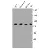
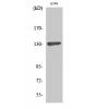
Fig1: Western blot analysis of Nfic on different cell lysate using anti-Nfic antibody at 1/500 dilution.
Positive control:
Lane 1: A549
Lane 2: SiHa
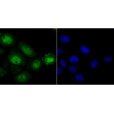
Fig2: ICC staining Nfic in A431 cells (green). The nuclear counter stain is DAPI (blue). Cells were fixed in paraformaldehyde, permeabilised with 0.25% Triton X100/PBS.
Fig3: ICC staining Nfic in A549 cells (green). The nuclear counter stain is DAPI (blue). Cells were fixed in paraformaldehyde, permeabilised with 0.25% Triton X100/PBS.
Fig4: ICC staining Nfic in SiHa cells (green). The nuclear counter stain is DAPI (blue). Cells were fixed in paraformaldehyde, permeabilised with 0.25% Triton X100/PBS.
Fig5: Immunohistochemical analysis of paraffin-embedded human liver tissue using anti-Nfic antibody. Counter stained with hematoxylin. The section was pre-treated using heat mediated antigen retrieval with sodium citrate buffer (pH6) for 20 mins.
Fig6: Flow cytometric analysis of SiHa cells with Nfic antibody at 1/100 dilution (purple) compared with an unlabelled control (cells without incubation with primary antibody; yellow). Alexa Fluor 488-conjugated goat anti-rabbit IgG was used as the secondary antibody.
特别提示:本公司的所有产品仅可用于科研实验,严禁用于临床医疗及其他非科研用途!