Anti-Rad21 antibody
-
概述
- 产品描述Cleavable component of the cohesin complex, involved in chromosome cohesion during cell cycle, in DNA repair, and in apoptosis. The cohesin complex is required for the cohesion of sister chromatids after DNA replication. The cohesin complex apparently forms a large proteinaceous ring within which sister chromatids can be trapped. At metaphase-anaphase transition, this protein is cleaved by separase/ESPL1 and dissociates from chromatin, allowing sister chromatids to segregate. The cohesin complex may also play a role in spindle pole assembly during mitosis. Also plays a role in apoptosis, via its cleavage by caspase-3/CASP3 or caspase-7/CASP7 during early steps of apoptosis: the C-terminal 64 kDa cleavage product may act as a nuclear signal to initiate cytoplasmic events involved in the apoptotic pathway.
- 产品名称Anti-Rad21 antibody
- 分子量120 kDa, predicted band size 72 kDa
- 种属反应性Human,Mouse,Rat
- 验证应用WB,ICC,IHC-P
- 抗体类型兔多抗
- 免疫原Recombinant protein within human Rad21 aa 250-440.
- 偶联Non-conjugated
-
性能
- 形态Liquid
- 浓度1 mg/ml.
- 存放说明Store at +4℃ after thawing. Aliquot store at -20℃. Avoid repeated freeze / thaw cycles.
- 存储缓冲液1*PBS (pH7.4), 0.2% BSA, 50% Glycerol. Preservative: 0.05% Sodium Azide.
- 亚型IgG
- 纯化方式Protein affinity purified.
- 亚细胞定位Centromere, Chromosome, Nucleus.
- 其它名称CDLS4 antibody
Double-strand-break repair protein rad21 homolog antibody
hHR21 antibody
HR21 antibody
HRAD21 antibody
KIAA0078 antibody
MCD1 antibody
Nuclear matrix protein 1 antibody
NXP-1 antibody
NXP1 antibody
Protein involved in DNA double-strand break repair antibody
RAD21 antibody
RAD21 homolog (S. pombe) antibody
RAD21 homolog antibody
RAD21_HUMAN antibody
Scc1 antibody
SCC1 homolog antibody
more
-
应用
WB:1:500-1:2,000
ICC:1:50-1:200
IHC-P:1:100-1:400
-
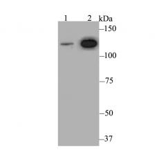
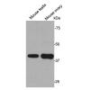
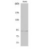
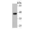
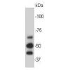
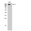
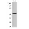
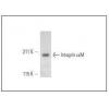
Fig1: Western blot analysis of Rad21 on different lysates. Proteins were transferred to a PVDF membrane and blocked with 5% BSA in PBS for 1 hour at room temperature. The primary antibody was used at a 1:500 dilution in 5% BSA at room temperature for 2 hours. Goat Anti-Rabbit IgG - HRP Secondary Antibody (HA1001) at 1:5,000 dilution was used for 1 hour at room temperature.
Positive control:
Lane 1: Mouse ovary tissue lysate
Lane 2: Daudi cell lysate
Fig2: ICC staining Rad21 in MG-63 cells (green). Formalin fixed cells were permeabilized with 0.1% Triton X-100 in TBS for 10 minutes at room temperature and blocked with 1% Blocker BSA for 15 minutes at room temperature. Cells were probed with Rad21 polyclonal antibody at a dilution of 1:100 for 1 hour at room temperature, washed with PBS. Alexa Fluorc™ 488 Goat anti-Rabbit IgG was used as the secondary antibody at 1/100 dilution. The nuclear counter stain is DAPI (blue).
Fig3: ICC staining Rad21 in SiHa cells (green). Formalin fixed cells were permeabilized with 0.1% Triton X-100 in TBS for 10 minutes at room temperature and blocked with 1% Blocker BSA for 15 minutes at room temperature. Cells were probed with Rad21 polyclonal antibody at a dilution of 1:100 for 1 hour at room temperature, washed with PBS. Alexa Fluorc™ 488 Goat anti-Rabbit IgG was used as the secondary antibody at 1/100 dilution. The nuclear counter stain is DAPI (blue).
Fig4: ICC staining Rad21 in SK-Br-3 cells (green). Formalin fixed cells were permeabilized with 0.1% Triton X-100 in TBS for 10 minutes at room temperature and blocked with 1% Blocker BSA for 15 minutes at room temperature. Cells were probed with Rad21 polyclonal antibody at a dilution of 1:100 for 1 hour at room temperature, washed with PBS. Alexa Fluorc™ 488 Goat anti-Rabbit IgG was used as the secondary antibody at 1/100 dilution. The nuclear counter stain is DAPI (blue).
Fig5: Immunohistochemical analysis of paraffin-embedded rat brain tissue using anti-Rad21 antibody. The section was pre-treated using heat mediated antigen retrieval with sodium citrate buffer (pH 6.0) for 20 minutes. The tissues were blocked in 5% BSA for 30 minutes at room temperature, washed with ddH2O and PBS, and then probed with the antibodyat 1/200 dilution, for 30 minutes at room temperature and detected using an HRP conjugated compact polymer system. DAB was used as the chrogen. Counter stained with hematoxylin and mounted with DPX.
Fig6: Immunohistochemical analysis of paraffin-embedded human thyroid gland cancer tissue using anti-Rad21 antibody. The section was pre-treated using heat mediated antigen retrieval with sodium citrate buffer (pH 6.0) for 20 minutes. The tissues were blocked in 5% BSA for 30 minutes at room temperature, washed with ddH2O and PBS, and then probed with the antibodyat 1/200 dilution, for 30 minutes at room temperature and detected using an HRP conjugated compact polymer system. DAB was used as the chrogen. Counter stained with hematoxylin and mounted with DPX.
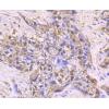
Fig7: Immunohistochemical analysis of paraffin-embedded human colon tissue using anti-Rad21 antibody. The section was pre-treated using heat mediated antigen retrieval with sodium citrate buffer (pH 6.0) for 20 minutes. The tissues were blocked in 5% BSA
Fig8: Immunohistochemical analysis of paraffin-embedded mouse testis tissue using anti-Rad21 antibody. The section was pre-treated using heat mediated antigen retrieval with sodium citrate buffer (pH 6.0) for 20 minutes. The tissues were blocked in 5% BSA
特别提示:本公司的所有产品仅可用于科研实验,严禁用于临床医疗及其他非科研用途!