


-

专业包装 正品保证
-

快乐服务 售后无忧
-

会员特权 优惠不断
-

个人信息 严格保护
| 货号 | 规格 | 可用库存 | 销售价(RMB) | 您的折扣价(RMB) | 购买数量 |
|---|
| 熔点: | |
|---|---|
| 密度: | |
| 储存条件: | -20℃ |
Anti-KV1.1 antibody
产品描述Voltage-gated potassium channel that mediates transmembrane potassium transport in excitable membranes, primarily in the brain and the central nervous system, but also in the kidney. Contributes to the regulation of the membrane potential and nerve signaling, and prevents neuronal hyperexcitability. Forms tetrameric potassium-selective channels through which potassium ions pass in accordance with their electrochemical gradient. The channel alternates between opened and closed conformations in response to the voltage difference across the membrane. Channel properties are modulated by cytoplasmic beta subunits that regulate the subcellular location of the alpha subunits and promote rapid inactivation of delayed rectifier potassium channels. In vivo, membranes probably contain a mixture of heteromeric potassium channel complexes, making it difficult to assign currents observed in intact tissues to any particular potassium channel family member. Regulates neuronal excitability in hippocampus, especially in mossy fibers and medial perforant path axons, preventing neuronal hyperexcitability. Response to toxins that are selective for KCNA1, respectively for KCNA2, suggests that heteromeric potassium channels composed of both KCNA1 and KCNA2 play a role in pacemaking and regulate the output of deep cerebellar nuclear neurons.
产品名称Anti-KV1.1 antibody
分子量56 kDa (Predicted band size)
种属反应性Human,Mouse,Rat
验证应用WB,IHC-P,FC
抗体类型兔多抗
免疫原Synthetic peptide within rat KV1.1 aa 190-250.
偶联Non-conjugated
形态Liquid
浓度1 mg/mL
存放说明Store at +4℃ after thawing. Aliquot store at -20℃. Avoid repeated freeze / thaw cycles.
存储缓冲液1*PBS (pH7.4), 0.2% BSA, 50% Glycerol. Preservative: 0.05% Sodium Azide.
亚型IgG
纯化方式Peptide affinity purified.
亚细胞定位Endoplasmic reticulum. Plasma membrane. Other locations.
其它名称
WB: 1:500-1:2,000
IHC-P: 1:50-1:200
FC: 1:50-1:100

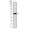
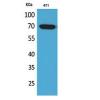
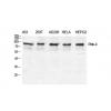
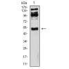
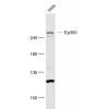
Fig1: Western blot analysis of KV1.1 on rat brain tissue lysate. Proteins were transferred to a PVDF membrane and blocked with 5% BSA in PBS for 1 hour at room temperature. The primary antibody was used in 5% BSA at room temperature for 2 hours. Goat Anti-Rabbit IgG - HRP Secondary Antibody (HA1001) at 1:5,000 dilution was used for 1 hour at room temperature.

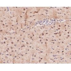
Fig2: Immunohistochemical analysis of paraffin-embedded rat spinal cord tissue using anti-KV1.1 antibody. The section was pre-treated using heat mediated antigen retrieval with Tris-EDTA buffer (pH 8.0-8.4) for 20 minutes.The tissues were blocked in 5% BSA for 30 minutes at room temperature, washed with ddH2O and PBS, and then probed with the primary antibody for 30 minutes at room temperature. The detection was performed using an HRP conjugated compact polymer system. DAB was used as the chromogen. Tissues were counterstained with hematoxylin and mounted with DPX.

Fig3: Immunohistochemical analysis of paraffin-embedded mouse brain tissue using anti-KV1.1 antibody. The section was pre-treated using heat mediated antigen retrieval with Tris-EDTA buffer (pH 8.0-8.4) for 20 minutes.The tissues were blocked in 5% BSA for 30 minutes at room temperature, washed with ddH2O and PBS, and then probed with the primary antibody for 30 minutes at room temperature. The detection was performed using an HRP conjugated compact polymer system. DAB was used as the chromogen. Tissues were counterstained with hematoxylin and mounted with DPX.

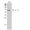
Fig4: Flow cytometric analysis of KV1.1 was done on A549 cells. The cells were fixed, permeabilized and stained with the primary antibody (red). After incubation of the primary antibody at room temperature for an hour, the cells were stained with a Alexa Fluor 488-conjugated Goat anti-Rabbit IgG Secondary antibody at 1/1000 dilution for 30 minutes.Unlabelled sample was used as a control (cells without incubation with primary antibody; black).
FC M
特别提示:本公司的所有产品仅可用于科研实验,严禁用于临床医疗及其他非科研用途!


