Anti-ITPR2 antibody
-
概述
- 产品描述The protein encoded by this gene belongs to the inositol 1,4,5-triphosphate receptor family, whose members are second messenger intracellular calcium release channels. These proteins mediate a rise in cytoplasmic calcium in response to receptor activated production of inositol triphosphate. Inositol triphosphate receptor-mediated signaling is involved in many processes including cell migration, cell division, smooth muscle contraction, and neuronal signaling. This protein is a type 2 receptor that consists of a cytoplasmic amino-terminus that binds inositol triphosphate, six membrane-spanning helices that contribute to the ion pore, and a short cytoplasmic carboxy-terminus. A mutation in this gene has been associated with anhidrosis, suggesting that intracellular calcium release mediated by this protein is required for eccrine sweat production.
- 产品名称Anti-ITPR2 antibody
- 分子量308 kDa
- 种属反应性Human,Mouse,Rat
- 验证应用Dot blot,ICC,FC,IHC-P
- 抗体类型兔多抗
- 免疫原Recombinant protein within human ITPR2 aa 1100-1450.
- 偶联Non-conjugated
-
性能
- 形态Liquid
- 浓度1 mg/mL.
- 存放说明Store at +4℃ after thawing. Aliquot store at -20℃. Avoid repeated freeze / thaw cycles.
- 存储缓冲液1*PBS (pH7.4), 0.2% BSA, 50% Glycerol. Preservative: 0.05% Sodium Azide.
- 亚型IgG
- 纯化方式Protein affinity purified.
- 亚细胞定位Endoplasmic reticulum membrane.
- 其它名称4 antibody
5-trisphosphate receptor antibody
Inositol 1 4 5 trisphosphate receptor type 2 antibody
Inositol 1,4,5-trisphosphate receptor type 2 antibody
Inositol 145 trisphosphate receptor type 2 antibody
InsP3 R2 antibody
InsP3R2 antibody
IP3 R2 antibody
IP3 receptor antibody
IP3 receptor isoform 2 antibody
IP3R 2 antibody
IP3R2 antibody
ITPR 2 antibody
Itpr2 antibody
ITPR2_HUMAN antibody
Type 2 inositol 1 4 5 trisphosphate receptor antibody
Type 2 inositol 1 antibody
Type 2 inositol 145 trisphosphate receptor antibody
Type 2 InsP3 receptor antibody
more
-
应用
Dot Blot: 1:2,000-1:5,000
ICC: 1:50-1:100
IHC-P: 1:50 -1:200
FC: 1:50-1:100
-
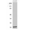
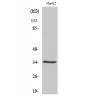
Fig1: Dot blot analysis of anti-ITPR2 on PVDF. 0-4ug antigens were given in this test. Anti-ITPR2 antibody was diluted with 1/500.
Fig2: ICC staining ITPR2 in HepG2 cells (green). The nuclear counter stain is DAPI (blue). Cells were fixed in paraformaldehyde, permeabilised with 0.25% Triton X100/PBS.
Fig3: ICC staining ITPR2 in HT-29 cells (green). The nuclear counter stain is DAPI (blue). Cells were fixed in paraformaldehyde, permeabilised with 0.25% Triton X100/PBS.
Fig4: ICC staining ITPR2 in SiHa cells (green). The nuclear counter stain is DAPI (blue). Cells were fixed in paraformaldehyde, permeabilised with 0.25% Triton X100/PBS.
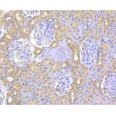
Fig5: Immunohistochemical analysis of paraffin-embedded rat kidney tissue using anti-ITPR2 antibody. Counter stained with hematoxylin.
Fig6: Immunohistochemical analysis of paraffin-embedded human colon tissue using anti-ITPR2 antibody. Counter stained with hematoxylin.
Fig7: Immunohistochemical analysis of paraffin-embedded human breast tissue using anti-ITPR2 antibody. Counter stained with hematoxylin.
Fig8: Immunohistochemical analysis of paraffin-embedded mouse liver tissue using anti-ITPR2 antibody. Counter stained with hematoxylin.
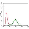
Fig9: Flow cytometric analysis of HepG2 cells with ITPR2 antibody at 1/100 dilution (red) compared with an unlabelled control (cells without incubation with primary antibody; blue). Alexa Fluor 488-conjugated goat anti-rabbit IgG was used as the secondary
特别提示:本公司的所有产品仅可用于科研实验,严禁用于临床医疗及其他非科研用途!