Anti-MMP9 antibody
-
概述
- 产品描述The matrix metalloproteinases (MMP) are a family of peptidase enzymes responsible for the degradation of extracellular matrix components, including collagen, gelatin, fibronectin, laminin and proteoglycan. Transcription of MMP genes is differentially activated by phorbol ester, lipopolysaccharide (LPS) or staphylococcal enterotoxin B (SEB). MMP catalysis requires both calcium and zinc. MMP-9 (also designated 92 kDa type IV collagenase or gelatinase B) has been shown to degrade bone collagens in concert with MMP-1 (also designated interstitial collagenase, fibroblast collagenase or collagenase-1), and cysteine proteases and may play a role in bone osteoclastic resorption. MMP-1 is downregulated by p53, and abnormality of p53 expression may contribute to joint degradation in rheumatoid arthritis by regulating MMP-1 expression.
- 产品名称Anti-MMP9 antibody
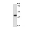
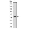
- 分子量100 kDa
- 种属反应性Human,Mouse
- 验证应用WB,ICC,IHC-P,FC
- 抗体类型兔多抗
- 免疫原Peptide
- 偶联Non-conjugated
-
性能
- 形态Liquid
- 浓度1 mg/mL.
- 存放说明Store at +4℃ after thawing. Aliquot store at -20℃ or -80℃. Avoid repeated freeze / thaw cycles.
- 存储缓冲液1*PBS (pH7.4), 0.2% BSA, 50% Glycerol. Preservative: 0.05% Sodium Azide.
- 亚型IgG
- 纯化方式Peptide affinity purified.
- 亚细胞定位Extracellular matrix. Secreted.
- 其它名称82 kDa matrix metalloproteinase-9 antibody
92 kDa gelatinase antibody
92 kDa type IV collagenase antibody
CLG 4B antibody
CLG4B antibody
Collagenase Type 4 beta antibody
Collagenase type IV 92 KD antibody
EC 3.4.24.35 antibody
Gelatinase 92 KD antibody
Gelatinase B antibody
Gelatinase beta antibody
GelatinaseB antibody
GELB antibody
Macrophage gelatinase antibody
MANDP2 antibody
Matrix metallopeptidase 9 (gelatinase B, 92kDa gelatinase, 92kDa type IV collagenase) antibody
Matrix Metalloproteinase 9 antibody
MMP 9 antibody
MMP-9 antibody
MMP9 antibody
MMP9_HUMAN antibody
Type V collagenase antibody
more
-
应用
ICC: 1:50-1:200
IHC-P: 1:50-1:200
FC: 1:50-1:100
WB: 1:500
-
Fig1: ICC staining MMP9 in Hela cells (green). The nuclear counter stain is DAPI (blue). Cells were fixed in paraformaldehyde, permeabilised with 0.25% Triton X100/PBS.
Fig2: ICC staining MMP9 in MCF-7 cells (green). The nuclear counter stain is DAPI (blue). Cells were fixed in paraformaldehyde, permeabilised with 0.25% Triton X100/PBS.
Fig3: ICC staining MMP9 in PANC-1 cells (green). The nuclear counter stain is DAPI (blue). Cells were fixed in paraformaldehyde, permeabilised with 0.25% Triton X100/PBS.
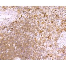
Fig4: Immunohistochemical analysis of paraffin-embedded human spleen tissue using anti-MMP9 antibody. Counter stained with hematoxylin.
Fig5: Immunohistochemical analysis of paraffin-embedded mouse spleen tissue using anti-MMP9 antibody. Counter stained with hematoxylin.
Fig6: Immunohistochemical analysis of paraffin-embedded human tonsil tissue using anti-MMP9 antibody. Counter stained with hematoxylin.
Fig7: Flow cytometric analysis of HL-60 cells with MMP9 antibody at 1/100 dilution (red) compared with an unlabelled control (cells without incubation with primary antibody; black). Goat anti rabbit IgG (FITC) was used as the secondary antibody
特别提示:本公司的所有产品仅可用于科研实验,严禁用于临床医疗及其他非科研用途!