Anti-NLRC3 antibody
-
概述
- 产品描述NLRC3, short for NOD-like receptor family CARD domain containing 3, is an intracellular protein that plays a role in the immune system. It was previously known as nucleotide-binding oligomerization domain, leucine rich repeat and CARD domain containing 3 (NOD3) and CLR16.2. NLRC3 inhibits the activity of T cells. Decreases the transcription of genes that are normally up-regulated after T-cell stimulation. Delays degradation of NFKBIA/IKBA.
- 产品名称Anti-NLRC3 antibody
- 分子量120 kDa
- 种属反应性Human,Mouse
- 验证应用WB,ICC,IHC-P,FC
- 抗体类型兔多抗
- 免疫原Peptide
- 偶联Non-conjugated
-
性能
- 形态Liquid
- 浓度1 mg/mL.
- 存放说明Store at +4℃ after thawing. Aliquot store at -20℃ or -80℃. Avoid repeated freeze / thaw cycles.
- 存储缓冲液1*PBS (pH7.4), 0.2% BSA, 50% Glycerol. Preservative: 0.05% Sodium Azide.
- 亚型IgG
- 纯化方式Peptide affinity purified.
- 亚细胞定位Cytoplasm. Cytoskeleton.
- 其它名称CARD15-like antibody
CARD15-like protein antibody
Caterpiller 16.2 antibody
Caterpiller protein 16.2 antibody
CLR16.2 antibody
FLJ00348 antibody
NLR family CARD domain containing 3 antibody
Nlrc3 antibody
NLRC3_HUMAN antibody
NOD-like receptor C3 antibody
NOD3 antibody
NOD3 protein antibody
Nucleotide-binding oligomerization domain protein 3 antibody
Nucleotide-binding oligomerization domain protein 3 caterpiller 16.2 antibody
nucleotide-binding oligomerization domain, leucine rich repeat and CARD domain containing 3 antibody
Protein NLRC3 antibody
more
-
应用
WB: 1:500-1:2000
ICC: 1:50-1:200
IHC-P: 1:50-1:200
FC: 1:50-1:200
-
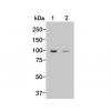
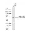
Fig1: Western blot analysis of NLRC3 on human thymus tissue lysates using anti-NLRC3 antibody at 1/500 dilution.
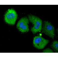
Fig2: ICC staining NLRC3 in HUVEC cells (green). The nuclear counter stain is DAPI (blue). Cells were fixed in paraformaldehyde, permeabilised with 0.25% Triton X100/PBS.
Fig3: ICC staining NLRC3 in MCF-7 cells (green). The nuclear counter stain is DAPI (blue). Cells were fixed in paraformaldehyde, permeabilised with 0.25% Triton X100/PBS.
Fig4: ICC staining NLRC3 in SH-SY5Y cells (green). The nuclear counter stain is DAPI (blue). Cells were fixed in paraformaldehyde, permeabilised with 0.25% Triton X100/PBS.
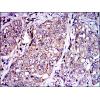
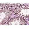
Fig5: Immunohistochemical analysis of paraffin-embedded human colon cancer tissue using anti-NLRC3 antibody. Counter stained with hematoxylin.
Fig6: Immunohistochemical analysis of paraffin-embedded human spleen tissue using anti-NLRC3 antibody. Counter stained with hematoxylin.
Fig7: Immunohistochemical analysis of paraffin-embedded human tonsil tissue using anti-NLRC3 antibody. Counter stained with hematoxylin.
Fig8: Immunohistochemical analysis of paraffin-embedded mouse colon tissue using anti-NLRC3 antibody. Counter stained with hematoxylin.
Fig9: Flow cytometric analysis of MCF-7 cells with NLRC3 antibody at 1/50 dilution (red) compared with an unlabelled control (cells without incubation with primary antibody; black). Alexa Fluor 488-conjugated Goat anti rabbit IgG was used as the secondar
特别提示:本公司的所有产品仅可用于科研实验,严禁用于临床医疗及其他非科研用途!