Anti-PKA C alpha antibody
-
概述
- 产品描述The second messenger cyclic AMP (cAMP) mediates diverse cellular responses to external signals such as proliferation, ion transport, regulation of metabolism and gene transcription by activation of the cAMP-dependent protein kinase (cAPK or PKA). Activation of PKA occurs when cAMP binds to the two regulatory subunits of the tetrameric PKA holoenzyme resulting in release of active catalytic subunits. Three catalytic (C) subunits have been identified, designated Cα, Cβ and Cγ, that each represent specific gene products. Cα and Cβ are closely related (93% amino acid sequence similarity), whereas Cγ displays 83% and 79% similarity to Cα and Cβ, respectively. Activation of transcription upon elevation of cAMP levels results from translocation of PKA to the nucleus where it phosphorylates the transcription factor cAMP response element binding protein (CREB) on serine 133 which in turn leads to TFIIB binding to TATA-box-binding protein TBP1, thus linking phospho-CREB to the pol II transcription initiation complex.
- 产品名称Anti-PKA C alpha antibody
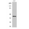
- 分子量40 kDa
- 种属反应性Human,Mouse,Rat
- 验证应用ICC,IHC-P,FC
- 抗体类型兔多抗
- 免疫原Recombinant protein.
- 偶联Non-conjugated
-
性能
- 形态Liquid
- 浓度1 mg/mL.
- 存放说明Store at +4℃ after thawing. Aliquot store at -20℃ or -80℃. Avoid repeated freeze / thaw cycles.
- 存储缓冲液1*PBS (pH7.4), 0.2% BSA, 50% Glycerol. Preservative: 0.05% Sodium Azide.
- 亚型IgG
- 纯化方式Protein affinity purified.
- 亚细胞定位Mitochondrion. Nucleus.Plasma membrane.
- 其它名称cAMP dependent protein kinase beta catalytic subunit antibody
cAMP dependent protein kinase alpha catalytic subunit antibody
cAMP dependent protein kinase catalytic subunit alpha antibody
cAMP dependent protein kinase catalytic subunit beta antibody
PKA C alpha antibody
PKA C beta antibody
PKACA antibody
PKACB antibody
PRKACA antibody
PRKACB antibody
Protein kinase cAMP dependent catalytic alpha antibody
Protein kinase cAMP dependent catalytic beta antibody
more
-
应用
ICC: 1:50-1:200
IHC-P: 1:50-1:200
FC: 1:50-1:100
-
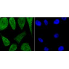
Fig1: ICC staining PKA C-alpha in A431 cells (green). The nuclear counter stain is DAPI (blue). Cells were fixed in paraformaldehyde, permeabilised with 0.25% Triton X100/PBS.
Fig2: ICC staining PKA C-alpha in PC-3M cells (green). The nuclear counter stain is DAPI (blue). Cells were fixed in paraformaldehyde, permeabilised with 0.25% Triton X100/PBS.
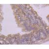
Fig3: Immunohistochemical analysis of paraffin-embedded rat brain tissue using anti-PKA C-alpha antibody. Counter stained with hematoxylin.
Fig4: Immunohistochemical analysis of paraffin-embedded human kidney tissue using anti-PKA C-alpha antibody. Counter stained with hematoxylin.
Fig5: Immunohistochemical analysis of paraffin-embedded mouse testis tissue using anti-PKA C-alpha antibody. Counter stained with hematoxylin.
Fig6: Flow cytometric analysis of PC-3M cells with PKA C-alpha antibody at 1/100 dilution (red) compared with an unlabelled control (cells without incubation with primary antibody; black
特别提示:本公司的所有产品仅可用于科研实验,严禁用于临床医疗及其他非科研用途!