Anti-IDO antibody
-
概述
- 产品描述Indoleamine 2,3-dioxygenase (IDO) is an IFN-γ inducible enzyme that catalyzes the degradation of the essential amino acid L-tryptophan to N-formylkynurenine. The gene encoding human IDO maps to chromosome 8p12-p11. IDO, also known as INDO, is an important modulator of immunological responses and protects allogeneic concepti from alloreactive maternal lymphocytes. IDO mediates an interesting inhibitory effect of HeLa cells co-cultured with human PBLs. The ILN-2-induced proliferation response of PBLs is diminished in the presence of HeLa cells while an IDO inhbitor negates this effect. Flow cytometric analysis indicates both mature and immature CD123-positive dentritic cells suppress T cell activity using IDO. IDO-transfected cells co-cultured with T cells reduces T cell proliferation. Additionally, adopted transfer of donor T cells reduces donor T cell numbers in IDO-transgenic mice. The pharmacological or genetic manipulation of IDO may be useful for supressing undesirable T cell response.
- 产品名称Anti-IDO antibody
- 分子量45 kDa
- 种属反应性Human,Mouse
- 验证应用WB,ICC,IHC-P,FC
- 抗体类型兔多抗
- 免疫原Recombinant protein
- 偶联Non-conjugated
-
性能
- 形态Liquid
- 浓度1 mg/mL.
- 存放说明Store at +4℃ after thawing. Aliquot store at -20℃ or -80℃. Avoid repeated freeze / thaw cycles.
- 存储缓冲液1*PBS (pH7.4), 0.2% BSA, 50% Glycerol. Preservative: 0.05% Sodium Azide.
- 亚型IgG
- 纯化方式Protein affinity purified
- 亚细胞定位Cytoplasm.
-
应用
WB: 1:500-1:2,000
ICC: 1:100-1:500
IHC-P: 1:50-1:200
FC: 1:50-1:100
-
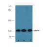
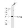
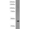
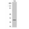
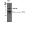
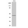
Fig1: Western blot analysis of IDO on human placenta tissue lysate using anti-IDO antibody at 1/500 dilution.
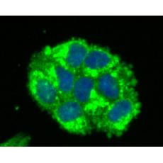
Fig2: ICC staining IDO in Hela cells (green). The nuclear counter stain is DAPI (blue). Cells were fixed in paraformaldehyde, permeabilised with 0.25% Triton X100/PBS.
Fig3: ICC staining IDO in HUVEC cells (green). The nuclear counter stain is DAPI (blue). Cells were fixed in paraformaldehyde, permeabilised with 0.25% Triton X100/PBS.
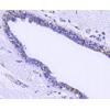
Fig4: Immunohistochemical analysis of paraffin-embedded human tonsil tissue using anti-IDO antibody. Counter stained with hematoxylin.
Fig5: Immunohistochemical analysis of paraffin-embedded human spleen tissue using anti-IDO antibody. Counter stained with hematoxylin.
Fig6: Flow cytometric analysis of HUVEC cells with IDO antibody at 1/100 dilution (red) compared with an unlabelled control (cells without incubation with primary antibody; black).
特别提示:本公司的所有产品仅可用于科研实验,严禁用于临床医疗及其他非科研用途!