Anti-GFAP antibody
-
概述
- 产品描述GFAP, a class-III intermediate filament, is a cell-specific marker that, during the development of the central nervous system, distinguishes astrocytes from other glial cells. In particular, vimentin filaments are present at early developmental stages, while GFAP filaments are characteristic of differentiated and mature brain astrocytes. In addition, GFAP intermediate filaments are also present in nonmyelin-forming Schwann cells in the peripheral nervous system.
- 产品名称Anti-GFAP antibody
- 分子量50 kDa
- 种属反应性Human,Mouse,Rat
- 验证应用WB,IHC-P,ICC,FC
- 抗体类型小鼠单抗
- 免疫原peptide
- 偶联Non-conjugated
-
性能
- 形态Liquid
- 浓度2 mg/mL.
- 存放说明Store at +4℃ after thawing. Aliquot store at -20℃ or -80℃. Avoid repeated freeze / thaw cycles.
- 存储缓冲液1*PBS (pH7.4), 0.2% BSA, 40% Glycerol. Preservative: 0.05% Sodium Azide.
- 亚型IgG1
- 纯化方式Protein A purified.
- 亚细胞定位Cytoplasm, intermediate filament
- 其它名称
- wu:fb34h11 antibody
- ALXDRD antibody
- cb345 antibody
more
-
应用
WB: 1:2,000-1:5,000
ICC:1:200
IHC-P: 1:200
FC: 1:200
-
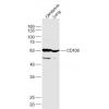
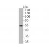
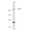
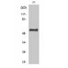
Fig1: Western blot analysis of GFAP on different cell lysates using anti- GFAP antibody at 1/5000 dilution.
Positive control: Line 1: Rat brain Line2 :Human brain
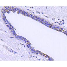
Fig2: Immunohistochemical analysis of paraffin-embedded mouse brain tissue using anti-GFAP antibody. Counter stained with hematoxylin.
Fig3: ICC staining of GFAP in A172 cells (red). Cells were fixed in paraformaldehyde, permeabilised with 0.25% Triton X100/PBS.
Fig4: ICC staining of GFAP in N2A cells (green). Cells were fixed in paraformaldehyde, permeabilised with 0.25% Triton X100/PBS.
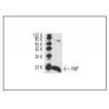
Fig5: Flow cytometric analysis of Hela cells with GFAP antibody at 1/100 dilution (blue) compared with an unlabelled control (cells without incubation with primary antibody; red). Goat anti mouse IgG (FITC) was used as the secondary antibody.
特别提示:本公司的所有产品仅可用于科研实验,严禁用于临床医疗及其他非科研用途!