Anti-Noggin antibody
-
概述
- 产品描述Genetic differentiation of the vertebrate somite necessitates a balance of inductive signals and antagonists. Noggin is a secreted protein that binds and inactivates members of the transforming growth factor-beta (TGFβ) superfamily of signaling proteins, such as bone morphogenetic proteins-2, 4, 7 (BMP2, BMP4, BMP7). Inhibition of BMP signaling by axially secreted Noggin mediates normal vertebrate skeletogenesis and patterning of the neural tube and somite. Spatially, Noggin may effectively antagonize BMP activity by efficiently diffusing through extracellular matrices, thereby creating morpho-genic gradients. Mice embryos that are homozygous null for Noggin, a lethal genotype, display stubby, continuous limbs with lack of joints in the paws and an array of other developmental defects.
- 产品名称Anti-Noggin antibody
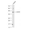
- 分子量26 kDa
- 种属反应性Human,Rat
- 验证应用WB,ICC,IHC-P,FC
- 抗体类型兔多抗
- 免疫原Recombinant protein
- 偶联Non-conjugated
-
性能
- 形态Liquid
- 浓度1 mg/mL.
- 存放说明Store at +4℃ after thawing. Aliquot store at -20℃ or -80℃. Avoid repeated freeze / thaw cycles.
- 存储缓冲液1*PBS (pH7.4), 0.2% BSA, 50% Glycerol. Preservative: 0.05% Sodium Azide.
- 亚型IgG
- 纯化方式Protein affinity purified
- 亚细胞定位Secreted.
- 其它名称Nog antibody
NOGG_HUMAN antibody
Noggin antibody
SYM 1 antibody
SYM1 antibody
Symphalangism 1 (proximal) antibody
Synostoses (multiple) syndrome 1 antibody
SYNS 1 antibody
SYNS1 antibody
more
-
应用
WB: 1:500
ICC: 1:50-1:200
IHC-P: 1:50-1:200
FC: 1:50-1:100
-
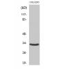
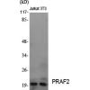
Fig1: Western blot analysis of Noggin on Recombinant protein using anti-Noggin antibody at 1/500 dilution.
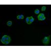
Fig2: ICC staining Noggin in PC-3M cells (green). The nuclear counter stain is DAPI (blue). Cells were fixed in paraformaldehyde, permeabilised with 0.25% Triton X100/PBS.
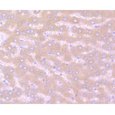
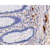
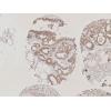
Fig3: Immunohistochemical analysis of paraffin-embedded human liver tissue using anti-Noggin antibody. Counter stained with hematoxylin.
Fig4: Immunohistochemical analysis of paraffin-embedded human placenta tissue using anti-Noggin antibody. Counter stained with hematoxylin.
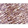
Fig5: Immunohistochemical analysis of paraffin-embedded rat liver tissue using anti-Noggin antibody. Counter stained with hematoxylin.
Fig6: Flow cytometric analysis of PC-3M cells with Noggin antibody at 1/100 dilution (red) compared with an unlabelled control (cells without incubation with primary antibody; black).
特别提示:本公司的所有产品仅可用于科研实验,严禁用于临床医疗及其他非科研用途!