Anti-NGF antibody
-
概述
- 产品描述Neurotrophins function to regulate naturally occurring cell death of neurons during development. The prototype neurotrophin is nerve growth factor (NGF), originally discovered in the 1950s as a soluble peptide promoting the survival of, and neurite outgrowth from, sympathetic ganglia. Three additional structurally homologous neurotrophic factors have been identified. These include brain-derived neurotrophic factor (BDNF), neurotrophin-3 (NT-3) and neurotrophin-4 (NT-4) (also designated NT-5). These various neurotrophins stimulate the in vitro survival of distinct, but partially overlapping, populations of neurons. The cell surface receptors through which neurotrophins mediate their activity have been identified. For instance, the Trk A receptor is the preferential receptor for NGF, but also binds NT-3 and NT-4. The Trk B receptor binds both BDNF and NT-4 equally well, and binds NT-3 to a lesser extent, while the Trk C receptor only binds NT-3.
- 产品名称Anti-NGF antibody
- 分子量27 kDa
- 种属反应性Human,Mouse,Rat
- 验证应用WB,ICC,IHC-P
- 抗体类型兔多抗
- 免疫原Synthetic peptide within Human NGF aa 100-150.
- 偶联Non-conjugated
-
性能
- 形态Liquid
- 浓度1 mg/mL.
- 存放说明Store at +4℃ after thawing. Aliquot store at -20℃. Avoid repeated freeze / thaw cycles.
- 存储缓冲液1*PBS (pH7.4), 0.2% BSA, 50% Glycerol. Preservative: 0.05% Sodium Azide.
- 亚型IgG
- 纯化方式Peptide affinity purified.
- 亚细胞定位Secreted.
- 其它名称Beta nerve growth factor antibody
Beta NGF antibody
Beta-nerve growth factor antibody
Beta-NGF antibody
HSAN5 antibody
MGC161426 antibody
MGC161428 antibody
Nerve growth factor (beta polypeptide) antibody
Nerve growth factor antibody
Nerve growth factor beta antibody
Nerve growth factor beta polypeptide antibody
Nerve growth factor beta subunit antibody
NGF antibody
NGF_HUMAN antibody
NGFB antibody
NID67 antibody
more
-
应用
WB: 1:500
ICC: 1:50-1:200
IHC-P: 1:50-1:200
-
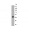
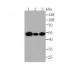
Fig1: Western blot analysis of NGF on recombinant protein lysate using anti-NGF antibody at 1/500 dilution.
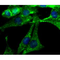
Fig2: ICC staining NGF in N2A cells (green). The nuclear counter stain is DAPI (blue). Cells were fixed in paraformaldehyde, permeabilised with 0.25% Triton X100/PBS.
Fig3: ICC staining NGF in SHG-44 cells (green). The nuclear counter stain is DAPI (blue). Cells were fixed in paraformaldehyde, permeabilised with 0.25% Triton X100/PBS.
Fig4: ICC staining NGF in SH-SY-5Y cells (green). The nuclear counter stain is DAPI (blue). Cells were fixed in paraformaldehyde, permeabilised with 0.25% Triton X100/PBS.
Fig5: Immunohistochemical analysis of paraffin-embedded rat brain tissue using anti-NGF antibody. Counter stained with hematoxylin.
Fig6: Immunohistochemical analysis of paraffin-embedded mouse brain tissue using anti-NGF antibody. Counter stained with hematoxylin.
特别提示:本公司的所有产品仅可用于科研实验,严禁用于临床医疗及其他非科研用途!