Anti-Rae1 antibody
-
概述
- 产品描述Mrnp41 (mRNA-binding protein, 41-KD), also known as Rae1 protein homolog and mRNA export factor, is a 368 amino acid protein that binds mRNA and is involved in nucleocytoplasmic transport. Though characterized in both the nucleus and cytoplasm, mrnp41 is primarily localized to the nuclear pore complex in the nuclear envelope. Mutations in mrnp41 may result in the accumulation of poly(A)-containing mRNA in the nucleus, further supporting the role of mrnp41 as a primary nuclear exporter of mRNA. Along with Nup98, mrnp41 has been shown to regulate E-cadherin, an activating subunit of the anaphase-promoting complex complex, which results in the prevention of securin degradation, therefore suggesting that mrnp41 may play a potential role in maintaining euploidy. Also, during mitosis, both mrnp41 and NuMA have been shown to be required for bipolar spindle formation.
- 产品名称Anti-Rae1 antibody
- 分子量41 kDa
- 种属反应性Human,Mouse,Rat
- 验证应用WB,ICC,FC
- 抗体类型兔多抗
- 免疫原Synthetic peptide within human Rae1 aa 330-368.
- 偶联Non-conjugated
-
性能
- 形态Liquid
- 浓度1 mg/mL.
- 存放说明Store at +4℃ after thawing. Aliquot store at -20℃. Avoid repeated freeze / thaw cycles.
- 存储缓冲液1*PBS (pH7.4), 0.2% BSA, 50% Glycerol. Preservative: 0.05% Sodium Azide.
- 亚型IgG
- 纯化方式Peptide affinity purified.
- 亚细胞定位Nucleus. Cytoskeleton.Cytoplasm.
- 其它名称dJ481F12.3 antibody
dJ800J21.1 antibody
FLJ30608 antibody
Homolog of yeast Rae1 (Bharathi) mRNA associated protein of 41 kDa (Kraemer) antibody
Homolog of yeast Rae1 mRNA associated protein of 41 kDa antibody
MGC117333 antibody
MGC126076 antibody
MGC126077 antibody
MIG 14 antibody
MIG14 antibody
Migration inducing gene 14 antibody
Mnrp 41 antibody
Mnrp41 antibody
mRNA associated protein mrnp 41 antibody
mRNA binding protein 41 kD antibody
mRNA export factor antibody
mRNA export protein antibody
mRNA-associated protein mrnp 41 antibody
MRNP 41 antibody
MRNP41 antibody
RAE 1 antibody
RAE1 (RNA export 1 S.pombe) homolog antibody
rae1 antibody
RAE1 homolog antibody
Rae1 protein homolog antibody
RAE1 RNA export 1 homolog (S. pombe) antibody
RAE1 RNA export 1 homolog antibody
RAE1L_HUMAN antibody
RNA export 1 antibody
RNA export 1 homolog antibody
more
-
应用
WB: 1:500-1,000
ICC: 1:50-1:200
FC: 1:50-1:200
-
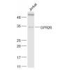
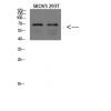
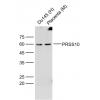
Fig1: Western blot analysis of Rae1 on different lysates using anti-Rae1 antibody at 1/500 dilution.
Positive control:
Lane 1: PC-3
Lane 2: Mouse testis tissue
Lane 3: Rat testis tissue
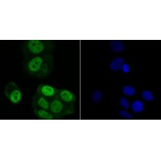
Fig2: ICC staining Rae1 in LOVO cells (green). The nuclear counter stain is DAPI (blue). Cells were fixed in paraformaldehyde, permeabilised with 0.25% Triton X100/PBS.
Fig3: ICC staining Rae1 in MCF-7 cells (green). The nuclear counter stain is DAPI (blue). Cells were fixed in paraformaldehyde, permeabilised with 0.25% Triton X100/PBS.
Fig4: ICC staining Rae1 in PC-3M cells (green). The nuclear counter stain is DAPI (blue). Cells were fixed in paraformaldehyde, permeabilised with 0.25% Triton X100/PBS.
Fig5: Flow cytometric analysis of LOVO cells with Rae1 antibody at 1/100 dilution (red) compared with an unlabelled control (cells without incubation with primary antibody; black). Alexa Fluor 488-conjugated goat anti-rabbit IgG was used as the secondary antibody.Flow cytometric analysis of LOVO cells with Rae1 antibody at 1/100 dilution (red) compared with an unlabelled control (cells without incubation with primary antibody; black). Alexa Fluor 488-conjugated goat anti-rabbit IgG was used as the secondary antibody.
特别提示:本公司的所有产品仅可用于科研实验,严禁用于临床医疗及其他非科研用途!