Anti-GST3 antibody
-
概述
- 产品描述Glutathione S-transferases (GSTs) function in the metabolic detoxification of various environmental carcinogens and lipid hydroperoxides. In response to oxidative stress, upregulation of the GST family member GSTP1 occurs, consistent with this function. Furthermore, the GSTP1 gene is subject to CpG island hypermethylation, a state that correlates with human prostatic carcinogenesis. GSTP1 gene hypermethylation can be detected in urine, ejaculate and plasma from men with prostate cancer, potentially making GSTP1 a useful biomarker for prostate cancer screening.
- 产品名称Anti-GST3 antibody
- 分子量23 kDa
- 种属反应性Human,Mouse,Rat
- 验证应用WB,ICC,IHC-P
- 抗体类型兔多抗
- 免疫原Recombinant protein with human GST3 aa 1-150.
- 偶联Non-conjugated
-
性能
- 形态Liquid
- 浓度1 mg/mL.
- 存放说明Store at +4℃ after thawing. Aliquot store at -20℃. Avoid repeated freeze / thaw cycles.
- 存储缓冲液1*PBS (pH7.4), 0.2% BSA, 50% Glycerol. Preservative: 0.05% Sodium Azide.
- 亚型IgG
- 纯化方式Protein affinity purified.
- 亚细胞定位Nucleus. Cytoplasm. Mitochondrion.
- 其它名称Deafness antibody
Deafness X-linked 7 antibody
DFN7 antibody
FAEES3 antibody
Fatty Acid Ethyl Ester Synthase III antibody
Glutathione S Transferase 3 antibody
Glutathione S Transferase Pi antibody
Glutathione S-transferase P antibody
Glutathione S-transferase pi 1 antibody
GST class-pi antibody
GST3 antibody
GSTP antibody
Gstp1 antibody
GSTP1-1 antibody
GSTP1_HUMAN antibody
PI antibody
X linked 7 antibody
more
-
应用
WB: 1:500-1:1,000
ICC: 1:500-1:1,000
IHC-P: 1:50-1:200
-
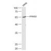
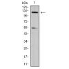
Fig1: Western blot analysis of GST3 on different cell lysate using anti-GST3 antibody at 1/1,000 dilution.
Positive control:
Lane 1: Siha
Lane 2: Hela
Lane 3: A549
Lane 4: Mouse kidney tissue
Lane 5: Rat liver tissue
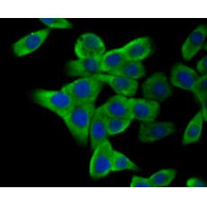
Fig2: ICC staining GST3 in A549 cells (green). The nuclear counter stain is DAPI (blue). Cells were fixed in paraformaldehyde, permeabilised with 0.25% Triton X100/PBS.
Fig3: ICC staining GST3 in HUVEC cells (green). The nuclear counter stain is DAPI (blue). Cells were fixed in paraformaldehyde, permeabilised with 0.25% Triton X100/PBS.
Fig4: ICC staining GST3 in LOVO cells (green). The nuclear counter stain is DAPI (blue). Cells were fixed in paraformaldehyde, permeabilised with 0.25% Triton X100/PBS.
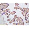
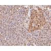
Fig5: Immunohistochemical analysis of paraffin-embedded human liver cancer tissue using anti-GST3 antibody. Counter stained with hematoxylin.
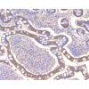
Fig6: Immunohistochemical analysis of paraffin-embedded human placenta tissue using anti-GST3 antibody. Counter stained with hematoxylin.
特别提示:本公司的所有产品仅可用于科研实验,严禁用于临床医疗及其他非科研用途!