


-

专业包装 正品保证
-

快乐服务 售后无忧
-

会员特权 优惠不断
-

个人信息 严格保护
| 货号 | 规格 | 可用库存 | 销售价(RMB) | 您的折扣价(RMB) | 购买数量 |
|---|
| 熔点: | |
|---|---|
| 密度: | |
| 储存条件: | -20℃ |
Anti-RYR2 antibody
产品描述Calcium channel that mediates the release of Ca2+ from the sarcoplasmic reticulum into the cytoplasm and thereby plays a key role in triggering cardiac muscle contraction. Aberrant channel activation can lead to cardiac arrhythmia. In cardiac myocytes, calcium release is triggered by increased Ca2+ levels due to activation of the L-type calcium channel CACNA1C. The calcium channel activity is modulated by formation of heterotetramers with RYR3. Required for cellular calcium ion homeostasis. Required for embryonic heart development.
产品名称Anti-RYR2 antibody
分子量565 kDa
种属反应性Human,Mouse,Rat
验证应用WB,ICC,IHC-P,FC
抗体类型兔多抗
免疫原Synthetic peptide within human RYR2 aa 1450-1550.
偶联Non-conjugated
形态Liquid
浓度1 mg/mL.
存放说明Store at +4℃ after thawing. Aliquot store at -20℃. Avoid repeated freeze / thaw cycles.
存储缓冲液1*PBS (pH7.4), 0.2% BSA, 50% Glycerol. Preservative: 0.05% Sodium Azide.
亚型IgG
纯化方式Peptide affinity purified.
亚细胞定位Membrane. Sarcoplasmic reticulum.
其它名称
WB: 1:500
ICC: 1:50-1:100
IHC-P: 1:50-1:200
FC: 1:50-1:100

Fig1: Dot blot analysis of anti-RYR2 immunization peptide on PVDF. 1ug, 2ug and 4ug peptides were given in this test. Anti-RYR2 antibody was diluted with 1/500.

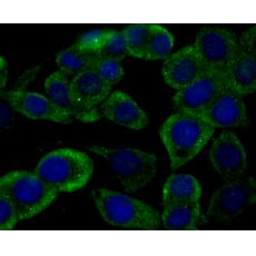
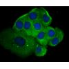
Fig2: ICC staining RYR2 in 293T cells (green). The nuclear counter stain is DAPI (blue). Cells were fixed in paraformaldehyde, permeabilised with 0.25% Triton X100/PBS.

Fig3: ICC staining RYR2 in LOVO cells (green). The nuclear counter stain is DAPI (blue). Cells were fixed in paraformaldehyde, permeabilised with 0.25% Triton X100/PBS.

Fig4: ICC staining RYR2 in SH-SY-5Y cells (green). The nuclear counter stain is DAPI (blue). Cells were fixed in paraformaldehyde, permeabilised with 0.25% Triton X100/PBS.

Fig5: Immunohistochemical analysis of paraffin-embedded rat skeletal muscle tissue using anti-RYR2 antibody. Counter stained with hematoxylin.

Fig6: Immunohistochemical analysis of paraffin-embedded human fetal skeletal muscle tissue using anti-RYR2 antibody. Counter stained with hematoxylin.

Fig7: Immunohistochemical analysis of paraffin-embedded mouse cerebellum tissue using anti-RYR2 antibody. Counter stained with hematoxylin.

Fig8: Flow cytometric analysis of 293T cells with RYR2 antibody at 1/100 dilution (fuchsia) compared with an unlabelled control (cells without incubation with primary antibody; yellow). Alexa Fluor 488-conjugated goat anti-rabbit IgG was used as the secon
特别提示:本公司的所有产品仅可用于科研实验,严禁用于临床医疗及其他非科研用途!


