


-

专业包装 正品保证
-

快乐服务 售后无忧
-

会员特权 优惠不断
-

个人信息 严格保护
| 货号 | 规格 | 可用库存 | 销售价(RMB) | 您的折扣价(RMB) | 购买数量 |
|---|
| 熔点: | |
|---|---|
| 密度: | |
| 储存条件: | -20℃ |
Anti-AMBP antibody
产品描述The AMBP (α-1-Microglobulin/bikunin precursor) gene encodes a protein precursor, known as AMBP, that is cleaved to produce two distinct proteins, designated a-1-Microglobulin and Bikunin. α-1-Microglobulin, also known as protein HC, is a member of the lipocalin superfamily and is secreted mainly in plasma, urine and cerebrospinal fluid. Thought to have reductase/dehydrogenase activity, α-1-Microglobulin exhibits immunosuppressive properties, such as cytokine secretion and inhibition of antigen-induced lymphocyte cell proliferation, and may be involved in the reduction of biological pro-oxidants. The second protein cleavage product, designated Bikunin and also known as inter-α-trypsin inhibitor light chain, ITI-LC or urinary trypsin inhibitor, is a widely expressed protein that is stored in the granules of human connective tissue mast cells. One of many proteins in the Kunitz-type protease inhibitor family, Bikunin prevents autodigestion by exocrine enzymes, such as trypsinogen and chymo-trypsinogen, and plays a role in the antiinflammatory/antiproteinase immune response. Unlike α-1-Microglobulin, Bikunin is implicated in the pathogenesis of a number of renal diseases, such as urolithiasis.
产品名称Anti-AMBP antibody
分子量39 kDa (Predicted band size)
种属反应性Human,Mouse
验证应用WB,ICC,IHC-P,FC
抗体类型兔多抗
免疫原Recombinant protein with Human AMBP aa 120-320.
偶联Non-conjugated
形态Liquid
浓度1 mg/mL.
存放说明Store at +4℃ after thawing. Aliquot store at -20℃. Avoid repeated freeze / thaw cycles.
存储缓冲液1*PBS (pH7.4), 0.2% BSA, 50% Glycerol. Preservative: 0.05% Sodium Azide.
亚型IgG
纯化方式Protein affinity purified.
亚细胞定位Secreted.
其它名称
WB: 1:500-1:1,000
ICC: 1:50-1:200
IHC-P: 1:50-1:200
FC: 1:50-1:100

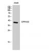
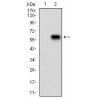
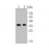
Fig1: Western blot analysis of AMBP on different cell lysates using anti-AMBP antibody at 1/1,000 dilution.
Positive control:
Lane 1: HepG2
Lane 2: Human liver
Lane 3: Mouse liver
Lane 4: Mouse brain

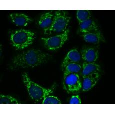
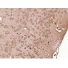
Fig2: ICC staining AMBP in HepG2 cells (green). The nuclear counter stain is DAPI (blue). Cells were fixed in paraformaldehyde, permeabilised with 0.25% Triton X100/PBS.

Fig3: ICC staining AMBP in MCF-7 cells (green). The nuclear counter stain is DAPI (blue). Cells were fixed in paraformaldehyde, permeabilised with 0.25% Triton X100/PBS.

Fig4: ICC staining AMBP in Hela cells (green). The nuclear counter stain is DAPI (blue). Cells were fixed in paraformaldehyde, permeabilised with 0.25% Triton X100/PBS.

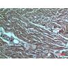
Fig5: Immunohistochemical analysis of paraffin-embedded human liver cancer tissue using anti-AMBP antibody. Counter stained with hematoxylin.

Fig6: Immunohistochemical analysis of paraffin-embedded human liver tissue using anti-AMBP antibody. Counter stained with hematoxylin.

Fig7: Immunohistochemical analysis of paraffin-embedded human kidney tissue using anti-AMBP antibody. Counter stained with hematoxylin.

Fig8: Flow cytometric analysis of HepG2 cells with AMBP antibody at 1/100 dilution (red) compared with an unlabelled control (cells without incubation with primary antibody; black). Alexa Fluor 488-conjugated goat anti-rabbit IgG was used as the secondary
特别提示:本公司的所有产品仅可用于科研实验,严禁用于临床医疗及其他非科研用途!


