


-

专业包装 正品保证
-

快乐服务 售后无忧
-

会员特权 优惠不断
-

个人信息 严格保护
| 货号 | 规格 | 可用库存 | 销售价(RMB) | 您的折扣价(RMB) | 购买数量 |
|---|
| 熔点: | |
|---|---|
| 密度: | |
| 储存条件: | -20℃ |
Anti-CDk1 antibody
产品描述Cdk1 is a small protein (approximately 34 kilodaltons), and is highly conserved. Cdk1 is comprised mostly by the bare protein kinase motif, which other protein kinases share. Cdk1, like other kinases, contains a cleft in which ATP fits. When bound to its cyclin partners, Cdk1 phosphorylation leads to cell cycle progression. Given its essential role in cell cycle progression, Cdk1 is highly regulated. Most obviously, Cdk1 is regulated by its binding with its cyclin partners. Cyclin binding alters access to the active site of Cdk1, allowing for Cdk1 activity; furthermore, cyclins impart specificity to Cdk1 activity. At least some cyclins contain a hydrophobic patch which may directly interact with substrates, conferring target specificity. Furthermore, cyclins can target Cdk1 to particular subcellular locations.
产品名称Anti-CDk1 antibody
分子量34 kDa
种属反应性Human,Mouse,Rat
验证应用WB,ICC,IHC-P,FC
抗体类型兔多抗
免疫原peptide
偶联Non-conjugated
形态Liquid
浓度1 mg/mL.
存放说明Store at +4℃ after thawing. Aliquot store at -20℃ or -80℃. Avoid repeated freeze / thaw cycles.
存储缓冲液1*PBS (pH7.4), 0.2% BSA, 40% Glycerol. Preservative: 0.05% Sodium Azide.
亚型IgG
纯化方式Peptide affinity purified
亚细胞定位Cytoplasm, nucleus
其它名称
WB: 1:500
ICC: 1:200
IHC-P: 1:200
FC: 1:100-1:200

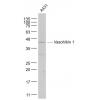
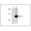
Fig1: Western blot analysis of CDk1 on different cell lysates using anti-CDk1 antibody at 1/500 dilution.
Positive control:
Lane 1: MCF-7
Lane 2: Jurkat
Lane 3: PC12
Lane 4: HepG2
Lane 5: Hela
Lane 6: NIH/3T3
Lane 7: Mouse liver
Lane 8: SKBR3

Fig2: ICC staining CDk1 in Hela cells (green). Cells were fixed in paraformaldehyde, permeabilised with 0.25% Triton X100/PBS.

Fig3: ICC staining CDk1 in HepG2 cells (green). Cells were fixed in paraformaldehyde, permeabilised with 0.25% Triton X100/PBS.

Fig4: ICC staining CDk1 in MCF-7 cells (green). Cells were fixed in paraformaldehyde, permeabilised with 0.25% Triton X100/PBS.

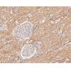
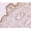
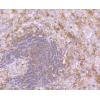
Fig5: Immunohistochemical analysis of paraffin-embedded rat spleen tissue using anti-CDk1 antibody. Counter stained with hematoxylin.

Fig6: Immunohistochemical analysis of paraffin-embedded human tonsil tissue using anti-CDk1 antibody. Counter stained with hematoxylin

Fig7: Immunohistochemical analysis of paraffin-e.mbedded human breast cancer tissue using anti-CDk1 antibody. Counter stained with hematoxylin

Fig8: Immunohistochemical analysis of paraffin-embedded mouse spleen tissue using anti-CDk1 antibody. Counter stained with hematoxylin.

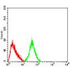
Fig9: Flow cytometric analysis of Hela cells with CDk1 antibody at 1/100 dilution (blue) compared with an unlabelled control (cells without incubation with primary antibody; red). Goat anti rabbit IgG (FITC) was used as the secondary antibody.
特别提示:本公司的所有产品仅可用于科研实验,严禁用于临床医疗及其他非科研用途!


