Anti-AKR1C1 antibody
-
概述
- 产品描述Human liver contains isoforms of dihydrodiol dehydrogenase (DD1, DD2, DD3 and DD4), which belong to the aldo-oxo reductase/aldo-keto reductase (AKR) superfamily, have 20α- or 3α-hydroxysteroid dehydrogenase (HSD) activity. DD1 is also designated AKR1C1, DDH or DDH1, while DD2 also can be designated AKR1C2, dDD, BABP or DDH2. AKR1C3 and 3α-HSD are alternate designations for human DD3 (which is referred to as AKR1C18 in rodents), while DD4 also can be called AKR1C4, CD, CHDR or AKR1C6 (in rodents). DD1 and DD2 are 20α-HSDs, whereas DD3 and DD4 are the 3α-HSDs. The multiple human cytosolic dihydrodiol dehydrogenases are involved in the metabolism of xenobiotics, such as polycyclic aromatic hydrocarbons, pesticides and steroid hormones, and are responsible for the reduction of ketone-containing drugs by using NADH or NADPH as a cofactor. The 20α-HSD catalyzes the reaction of progesterone to the inactive form 20α-hydroxyprogesterone. The 3α-HSD is a cytosolic, monomeric, NADPH-dependent oxidoreductase that reduces 3-keto-5-dihydrosteroids to their tetrahydro products. DD1 and DD2 are ubiquitously expressed, whereas DD4 mRNA is restricted to the liver. DD3 is a unique enzyme that can specifically catalyze the dehydrogenation of trans-benzenedihydrodiol and trans-naphthalenedihydrodiol.
- 产品名称Anti-AKR1C1 antibody
- 分子量37 kDa
- 种属反应性Human,Mouse
- 验证应用WB,IHC-P,FC
- 抗体类型兔多抗
- 免疫原Recombinant protein
- 偶联Non-conjugated
-
性能
- 形态Liquid
- 浓度1 mg/mL.
- 存放说明Store at +4℃ after thawing. Aliquot store at -20℃ or -80℃. Avoid repeated freeze / thaw cycles.
- 存储缓冲液1*PBS (pH7.4), 0.2% BSA, 40% Glycerol. Preservative: 0.05% Sodium Azide.
- 亚型IgG
- 纯化方式Protein A purified.
- 亚细胞定位Cytoplasm.
- 其它名称
- 2-dihydrobenzene-1 antibody
- 2-diol dehydrogenase antibody
- 20 alpha (3 alpha) hydroxysteroid dehydrogenase antibody 20 ALPHA HSD antibody
more
-
应用
WB: 1:500-1:2,000
IHC-P: 1:50-1:200
FC: 1:50-1:200
-
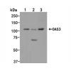
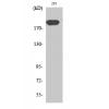
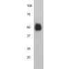
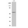
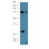
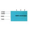
Fig1: Western blot analysis of AKR1C1 on different lysate using anti-AKR1C1 antibody at 1/1,000 dilution.
Positive control:
Lane 1: Mouse testis
Lane 2: SW480
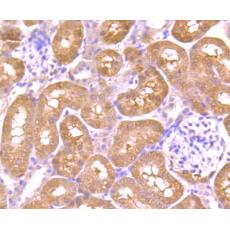
Fig2: Immunohistochemical analysis of paraffin-embedded human liver cancer tissue using anti-AKR1C1 antibody. Counter stained with hematoxylin.
Fig3: Immunohistochemical analysis of paraffin-embedded mouse kidney tissue using anti-AKR1C1 antibody. Counter stained with hematoxylin.
Fig4: Immunohistochemical analysis of paraffin-embedded mouse stomach tissue using anti-AKR1C1 antibody. Counter stained with hematoxylin.
Fig5: Flow cytometric analysis of HepG2 cells with AKR1C1 antibody at 1/100 dilution (blue) compared with an unlabelled control (cells without incubation with primary antibody; red).
特别提示:本公司的所有产品仅可用于科研实验,严禁用于临床医疗及其他非科研用途!