Anti-Cathepsin D antibody
-
概述
- 产品描述The cathepsin family of proteolytic enzymes contains several diverse classes of proteases. The cysteine protease class comprises cathepsins B, L, H, K, S, and O. The aspartyl protease class is composed of cathepsins D and E. Cathepsin G is in the serine protease class. Most cathepsins are lysosomal and each is involved in cellular metabolism, participating in various events such as peptide biosynthesis and protein degradation. Cathepsins may also cleave some protein precursors, thereby releasing regulatory peptides. The promoter region of the cathepsin D gene contains five Sp1 binding sites and four AP-2 binding sites.
- 产品名称Anti-Cathepsin D antibody
- 分子量27 kDa
- 种属反应性Human,Mouse
- 验证应用WB,ICC,IHC-P,FC
- 抗体类型兔多抗
- 免疫原Peptide
- 偶联Non-conjugated
-
性能
- 形态Liquid
- 浓度1 mg/mL.
- 存放说明Store at +4℃ after thawing. Aliquot store at -20℃ or -80℃. Avoid repeated freeze / thaw cycles.
- 存储缓冲液1*PBS (pH7.4), 0.2% BSA, 50% Glycerol. Preservative: 0.05% Sodium Azide.
- 亚型IgG
- 纯化方式Peptide affinity purified
- 亚细胞定位Lysosome. Melanosome. Secreted, extracellular space.
- 其它名称
- CatD antibody
- CATD_HUMAN antibody
- Cathepsin D antibody
more
-
应用
WB: 1:500
ICC: 1:50-1:200
IHC-P: 1:50-1:200
FC: 1:50-1:200
-
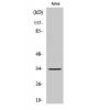
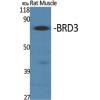
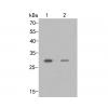
Fig1: Western blot analysis of Cathepsin D on MCF-7 cell lysate using anti-Cathepsin D antibody at 1/1,000 dilution.
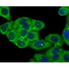
Fig2: ICC staining Cathepsin D in Hela cells (green). The nuclear counter stain is DAPI (blue). Cells were fixed in paraformaldehyde, permeabilised with 0.25% Triton X100/PBS.
Fig3: ICC staining Cathepsin D in HepG2 cells (green). The nuclear counter stain is DAPI (blue). Cells were fixed in paraformaldehyde, permeabilised with 0.25% Triton X100/PBS.
Fig4: ICC staining Cathepsin D in MCF-7 cells (green). The nuclear counter stain is DAPI (blue). Cells were fixed in paraformaldehyde, permeabilised with 0.25% Triton X100/PBS.
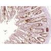
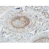
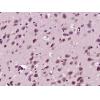
Fig5: Immunohistochemical analysis of paraffin-embedded human lung tissue using anti-Cathepsin D antibody. Counter stained with hematoxylin.
Fig6: Immunohistochemical analysis of paraffin-embedded human liver tissue using anti-Cathepsin D antibody. Counter stained with hematoxylin.
Fig7: Flow cytometric analysis of HepG2 cells with Cathepsin D antibody at 1/100 dilution (red) compared with an unlabelled control (cells without incubation with primary antibody; black).
Fig8: Flow cytometric analysis of MCF-7 cells with Cathepsin D antibody at 1/100 dilution (red) compared with an unlabelled control (cells without incubation with primary antibody; black).
特别提示:本公司的所有产品仅可用于科研实验,严禁用于临床医疗及其他非科研用途!