Anti-eIF-6 antibody
-
概述
- 产品描述Binds to the 60S ribosomal subunit and prevents its association with the 40S ribosomal subunit to form the 80S initiation complex in the cytoplasm. Behaves as a stimulatory translation initiation factor downstream insulin/growth factors. Is also involved in ribosome biogenesis. Associates with pre-60S subunits in the nucleus and is involved in its nuclear export. Cytoplasmic release of TIF6 from 60S subunits and nuclear relocalization is promoted by a RACK1 (RACK1)-dependent protein kinase C activity. In tissues responsive to insulin, controls fatty acid synthesis and glycolysis by exerting translational control of adipogenic transcription factors such as CEBPB, CEBPD and ATF4 that have G/C rich or uORF in their 5'UTR. Required for ROS-dependent megakaryocyte maturation and platelets formation, controls the expression of mitochondrial respiratory chain genes involved in reactive oxygen species (ROS) synthesis (By similarity). Involved in miRNA-mediated gene silencing by the RNA-induced silencing complex (RISC). Required for both miRNA-mediated translational repression and miRNA-mediated cleavage of complementary mRNAs by RISC . Modulates cell cycle progression and global translation of pre-B cells, its activation seems to be rate-limiting in tumorigenesis and tumor growth (By similarity).
- 产品名称Anti-eIF-6 antibody
- 分子量26 kDa
- 种属反应性Human,Mouse
- 验证应用WB,IHC,FC
- 抗体类型兔多抗
- 免疫原Recombinant protein within human eIF-6 aa 1-200.
- 偶联Non-conjugated
-
性能
- 形态Liquid
- 浓度1 mg/ml.
- 存放说明Store at +4℃ after thawing. Aliquot store at -20℃. Avoid repeated freeze / thaw cycles.
- 存储缓冲液1*TBS (pH7.4), 0.2% BSA, 50% Glycerol. Preservative: 0.05% Sodium Azide.
- 亚型IgG
- 纯化方式Protein affinity purified.
- 亚细胞定位Centromere, Chromosome, Nucleus.
- 其它名称
- b(2)gcn antibody
- B(2)GCN homolog antibody
- B4 integrin interactor antibody
more
-
应用
WB: 1:500-1:1,000
IHC: 1:500-1:2,000
FC: 1:50-1:100
-
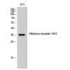
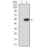
Fig1: Western blot analysis of eIF-6 on different lysates. Proteins were transferred to a PVDF membrane and blocked with 5% BSA in PBS for 1 hour at room temperature. The primary antibody was used in 5% BSA at room temperature for 2 hours. Goat Anti-Rabbit IgG - HRP Secondary Antibody (HA1001) at 1:5,000 dilution was used for 1 hour at room temperature.
Positive control:
Lane 1: SHSY5Y cell lysate
Lane 2: A549 cell lysate
Lane 3: HepG2 cell lysate
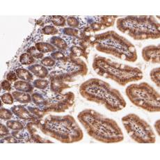
Fig2: Immunohistochemical analysis of paraffin-embedded human colon tissue using anti-eIF-6 antibody. The section was pre-treated using heat mediated antigen retrieval with sodium citrate buffer (pH 6.0) for 20 minutes. The tissues were blocked in 5% BSA for 30 minutes at room temperature, washed with ddH2O and PBS, and then probed with the primary antibody for 30 minutes at room temperature. The detection was performed using an HRP conjugated compact polymer system. DAB was used as the chromogen. Tissues were counterstained with hematoxylin and mounted with DPX.
Fig3: Immunohistochemical analysis of paraffin-embedded mouse colon tissue using anti-eIF-6 antibody. The section was pre-treated using heat mediated antigen retrieval with sodium citrate buffer (pH 6.0) for 20 minutes. The tissues were blocked in 5% BSA for 30 minutes at room temperature, washed with ddH2O and PBS, and then probed with the primary antibodyfor 30 minutes at room temperature. The detection was performed using an HRP conjugated compact polymer system. DAB was used as the chromogen. Tissues were counterstained with hematoxylin and mounted with DPX.
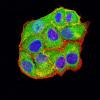
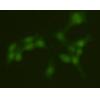
Fig4: Flow cytometric analysis of eIF-6 was done on SW620 cells. The cells were fixed, permeabilized and stained with the primary antibody (red). After incubation of the primary antibody at room temperature for an hour, the cells were stained with a Alexa Fluor 488-conjugated Goat anti-Rabbit IgG Secondary antibody at 1/1000 dilution for 30 minutes.Unlabelled sample was used as a control (cells without incubation with primary antibody; black).
特别提示:本公司的所有产品仅可用于科研实验,严禁用于临床医疗及其他非科研用途!