Anti-TCF4 antibody
-
概述
- 产品描述TCF7L2 is a transcription factor influencing the transcription of several genes thereby exerting a large variety of functions within the cell. It is a member of the Wnt signaling pathway. Stimulation of the pathway leads to the association of β-catenin with BCL9, translocation to the nucleus, and association with TCF7L2, which in turn results in the activation of Wnt target genes, specifically repressing proglucagon synthesis in enteroendocrine cells. TCF7L2 is implicated in a large variety of diseases. Several single nucleotide polymorphisms are associated with type 2 diabetes. In European populations it was found to be a major determinant of type 2 risk. A frameshift mutation of TCF7L2 is implicated in colorectal cancer .Variants of the gene are most likely involved in many other cancer types
- 产品名称Anti-TCF4 antibody
- 分子量40-80 kDa (different isoforms)
- 种属反应性Human,Mouse,Rat
- 验证应用WB,IHC-P,ICC,FC
- 抗体类型兔多抗
- 免疫原Synthetic peptide (KLH-coupled) within human TCF4 N-terminal.
- 偶联Non-conjugated
-
性能
- 形态Liquid
- 浓度1 mg/mL.
- 存放说明Store at +4℃ after thawing. Aliquot store at -20℃. Avoid repeated freeze / thaw cycles.
- 存储缓冲液1*PBS (pH7.4), 0.2% BSA, 40% Glycerol. Preservative: 0.05% Sodium Azide.
- 亚型IgG
- 纯化方式Peptide affinity purified
- 亚细胞定位Nucleus
- 其它名称
- bHLHb19 antibody
- Class B basic helix-loop-helix protein 19 antibody
- E2 2 antibody
more
-
应用
WB: 1:1,000-1:2,000
ICC: 1:500-1:2,000
IHC-P: 1:100-1:200
FC: 1:50-1:100
-
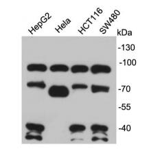
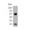
Fig1: Western blot analysis of TCF4 on different cell lysates using anti-TCF4 antibody at 1/1,000 dilution.
Fig2: ICC staining TCF4 in Hela cells (green). The nuclear counter stain is DAPI (blue). Cells were fixed in paraformaldehyde, permeabilised with 0.25% Triton X100/PBS.
Fig3: ICC staining TCF4 in HepG2 cells (green). The nuclear counter stain is DAPI (blue). Cells were fixed in paraformaldehyde, permeabilised with 0.25% Triton X100/PBS.
Fig4: Immunohistochemical analysis of paraffin-embedded rat kidney tissue using anti-TCF4 antibody. Counter stained with hematoxylin.
Fig5: Immunohistochemical analysis of paraffin-embedded human tonsil tissue using anti-TCF4 antibody. Counter stained with hematoxylin.
Fig6: Immunohistochemical analysis of paraffin-embedded human colon cancer tissue using anti-TCF4 antibody. Counter stained with hematoxylin.
Fig7: Immunohistochemical analysis of paraffin-embedded human breast cancer tissue using anti-TCF4 antibody. Counter stained with hematoxylin.
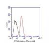
Fig8: Flow cytometric analysis of HepG2 cells with TCF4 antibody at 1/100 dilution (red) compared with an unlabelled control (cells without incubation with primary antibody; black). Alexa Fluor 488-conjugated goat anti-rabbit IgG was used as the secondary
特别提示:本公司的所有产品仅可用于科研实验,严禁用于临床医疗及其他非科研用途!