Anti-EEA1 antibody
-
概述
- 产品描述EEA1 localizes exclusively to early endosomes and has an important role in endosomal trafficking. EEA1 binds directly to the phospholipid phosphatidylinositol 3-phosphate through its C-terminal FYVE domain and forms a homodimer through a coiled coil . EEA1 acts as a tethering molecule that couples vesicle docking with SNAREs such as N-ethylmaleimide sensitive fusion protein, bringing the endosomes physically closer and ultimately resulting in the fusion and delivery of endosomal cargo. Due to the proteins importance in vesicular trafficking, a number of intracellular bacteria prevent EEA1 recruitment to the vacuole. Mycobacterium tuberculosis is known to inhibit the recruitment of EEA1 to the phagosomal membrane through CamKII.
- 产品名称Anti-EEA1 antibody
- 分子量162 kDa
- 种属反应性Human,Mouse,Rat
- 验证应用WB,ICC,IHC-P,FC
- 抗体类型兔多抗
- 免疫原Synthetic peptide of the N terminal residues of Human EEA1.
- 偶联Non-conjugated
-
性能
- 形态Liquid
- 浓度1 mg/mL.
- 存放说明Store at +4℃ after thawing. Aliquot store at -20℃. Avoid repeated freeze / thaw cycles.
- 存储缓冲液1*PBS (pH7.4), 0.2% BSA, 40% Glycerol. Preservative: 0.05% Sodium Azide.
- 亚型IgG
- 纯化方式Peptide affinity purified.
- 亚细胞定位Cytoplasm, early endosome membrane.
- 其它名称
- Early endosome antigen 1 antibody
- Early endosome antigen 1, 162kD antibody
- Early endosome associated protein antibody
more
-
应用
WB:1:1,000
ICC:1:100-1:200
IHC-P:1:50-1:200
FC:1:50-1:100
-
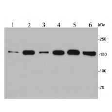
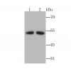
Fig1: Western blot analysis of EEA1 on different lysates. Proteins were transferred to a PVDF membrane and blocked with 5% BSA in PBS for 1 hour at room temperature. The primary antibody was used at a 1:500 dilution in 5% BSA at room temperature for 2 hours. Goat Anti-Rabbit IgG - HRP Secondary Antibody (HA1001) at 1:5,000 dilution was used for 1 hour at room temperature.
Positive control:
Lane 1: NIH/3T3 cell lysate
Lane 2: Hela cell lysate
Lane 3: Jurkat cell lysate
Lane 4: 293T cell lysate
Lane 5: A431 cell lysate
Lane 6: Rat brain tissue lysate
Fig2: ICC staining EEA1 in NIH/3T3 cells (green). Formalin fixed cells were permeabilized with 0.1% Triton X-100 in TBS for 10 minutes at room temperature and blocked with 1% Blocker BSA for 15 minutes at room temperature. Cells were probed with the antibody (R1401-23) at a dilution of 1:100 for 1 hour at room temperature, washed with PBS. Alexa Fluorc™ 488 Goat anti-Rabbit IgG was used as the secondary antibody at 1/100 dilution.
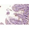
Fig3: Immunohistochemical analysis of paraffin-embedded rat prostate tissue using anti-EEA1 antibody. The section was pre-treated using heat mediated antigen retrieval with Tris-EDTA buffer (pH 8.0-8.4) for 20 minutes.The tissues were blocked in 5% BSA for 30 minutes at room temperature, washed with ddH2O and PBS, and then probed with the antibody (R1401-23) at 1/50 dilution, for 30 minutes at room temperature and detected using an HRP conjugated compact polymer system. DAB was used as the chrogen. Counter stained with hematoxylin and mounted with DPX.
Fig4: Immunohistochemical analysis of paraffin-embedded mouse colon tissue using anti-EEA1 antibody. The section was pre-treated using heat mediated antigen retrieval with Tris-EDTA buffer (pH 8.0-8.4) for 20 minutes.The tissues were blocked in 5% BSA for 30 minutes at room temperature, washed with ddH2O and PBS, and then probed with the antibody (R1401-23) at 1/50 dilution, for 30 minutes at room temperature and detected using an HRP conjugated compact polymer system. DAB was used as the chrogen. Counter stained with hematoxylin and mounted with DPX.
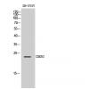
Fig5: Flow cytometric analysis of EEA1 was done on Hela cells. The cells were fixed, permeabilized and stained with EEA1 antibody at 1/100 dilution (red) compared with an unlabelled control (cells without incubation with primary antibody; black). After incubation of the primary antibody on room temperature for an hour, the cells was stained with a Alexa Fluor™ 488-conjugated goat anti-rabbit IgG Secondary antibody at 1/500 dilution.
特别提示:本公司的所有产品仅可用于科研实验,严禁用于临床医疗及其他非科研用途!