Anti-WDR5 antibody
-
概述
- 产品描述WD-repeat protein 5 (WDR5, also designated BMP-2-induced gene 3 kb or BIG-3) belongs to the family of WD-40 repeat proteins, and is essential for vertebrate development, Hox gene activation and global H3K4 trimethylation. WDR5 is a conserved subunit of Trithorax (TRX) histone methyltransferase complexes that selectively binds to dimethylated Lys4 (K4me2) in histone H3 to promote K4 trimethylation by TRX. It is expressed in osteoblasts, chondrocytes, osteocytes and marrow stromal cells. The WDR5 protein contains 7 WD-repeats, which may play a role in its function of accelerating osteoblast differentiation.
- 产品名称Anti-WDR5 antibody
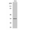
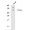
- 分子量36 kDa, additional band 70 kDa.
- 种属反应性Human
- 验证应用WB,ICC,FC
- 抗体类型兔多抗
- 免疫原Recombinant protein
- 偶联Non-conjugated
-
性能
- 形态Liquid
- 浓度1 mg/mL.
- 存放说明Store at +4℃ after thawing. Aliquot store at -20℃ or -80℃. Avoid repeated freeze / thaw cycles.
- 存储缓冲液1*PBS (pH7.4), 0.2% BSA, 50% Glycerol. Preservative: 0.05% Sodium Azide.
- 亚型IgG
- 纯化方式Protein affinity purified.
- 亚细胞定位Nucleus.
- 其它名称
- 2410008O07Rik antibody
- AA408785 antibody
- AA960360 antibody
more
-
应用
WB: 1:500-1:1,000
ICC: 1:50-1:200
FC: 1:50-1:100
-
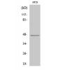
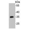
Fig1: Western blot analysis of WDR5 on MCF-7 cell lysate using anti-WDR5 antibody at 1/500 dilution.
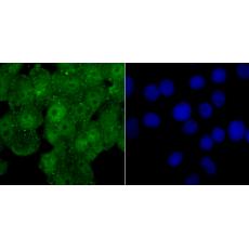
Fig2: ICC staining WDR5 in Hela cells (green). The nuclear counter stain is DAPI (blue). Cells were fixed in paraformaldehyde, permeabilised with 0.25% Triton X100/PBS.
Fig3: ICC staining WDR5 in MCF-7 cells (green). The nuclear counter stain is DAPI (blue). Cells were fixed in paraformaldehyde, permeabilised with 0.25% Triton X100/PBS.
Fig4: ICC staining WDR5 in PC-3M cells (green). The nuclear counter stain is DAPI (blue). Cells were fixed in paraformaldehyde, permeabilised with 0.25% Triton X100/PBS.
Fig5: Flow cytometric analysis of MCF-7 cells with WDR5 antibody at 1/100 dilution (red) compared with an unlabelled control (cells without incubation with primary antibody; black). Alexa Fluor 488-conjugated goat anti rabbit IgG was used as the secondary antibody.
特别提示:本公司的所有产品仅可用于科研实验,严禁用于临床医疗及其他非科研用途!