Anti-Thymidine Phosphorylase antibody
-
概述
- 产品描述Recent research has found that thymidine phosphorylase is also involved in angiogenesis. Experiments show inhibition of angiogenic effect by thymidine phosphorylase in the presence of 6-amino-5-chlorouracil, an inhibitor of thymidine phosphorylase, suggesting that the enzymatic activity of thymidine phosphorylase is required for its angiogenic activity. Thymidine phosphorylase has been determined to be almost identical to the platelet-derived endothelial cell growth factor (PD-ECGF). Although the mechanism of angiogenesis by thymidine phosphorylase is not yet known, reports show that the enzyme itself is not a growth factor but indirectly causes angiogenesis by stimulating chemotaxis of endothelial and other cells. Some reports suggest that thymidine phosphorylase promotes endothelial cell growth by reducing levels of thymidine that would otherwise inhibit endothelial cell growth. An alternative explanation is that the enzyme’s products induce angiogenesis. Experiments have found that 2-deoxyribose is an endothelial-cell chemoattractant and angiogenesis-inducing factor, which supports this explanation.Research has found thymidine phosphorylase is involved in angiogenesis during the menstrual cycle. The enzyme's expression in the endometrium is raised by a combination of progesterone and transforming growth factor-β1 and varies over the course of the menstrual cycle.
- 产品名称Anti-Thymidine Phosphorylase antibody
- 分子量Predicted band size 50 kDa.
- 种属反应性Human
- 验证应用WB,IHC-P,FC
- 抗体类型兔多抗
- 免疫原Recombinant protein within corresponding to N terminal of Human Thymidine Phosphorylase.
- 偶联Non-conjugated
-
性能
- 形态Liquid
- 浓度1 mg/ml.
- 存放说明Store at +4℃ after thawing. Aliquot store at -20℃. Avoid repeated freeze / thaw cycles.
- 存储缓冲液1*PBS (pH7.4), 0.2% BSA, 50% Glycerol. Preservative: 0.05% Sodium Azide.
- 亚型IgG
- 纯化方式Protein affinity purified.
- 亚细胞定位Cytosol.
- 其它名称
- ECGF 1 antibody
- ECGF antibody
- ECGF1 antibody
more
-
应用
WB: 1:500-1:2,000
IHC-P: 1:50-1:200
FC:1:100-1:200
-
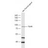
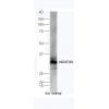
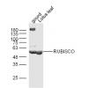
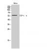
Fig1: Western blot analysis of Thymidine Phosphorylase on SKBR-3 cell lysate. Proteins were transferred to a PVDF membrane and blocked with 5% BSA in PBS for 1 hour at room temperature. The primary antibody was used in 5% BSA at room temperature for 2 hours. Goat Anti-Rabbit IgG - HRP Secondary Antibody (HA1001) at 1:5,000 dilution was used for 1 hour at room temperature.
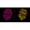
Fig2: Immunohistochemical analysis of paraffin-embedded human appendix tissue using anti-Thymidine Phosphorylase antibody. The section was pre-treated using heat mediated antigen retrieval with Tris-EDTA buffer (pH 8.0-8.4) for 20 minutes.The tissues were blocked in 5% BSA for 30 minutes at room temperature, washed with ddH2O and PBS, and then probed with the primary antibody for 30 minutes at room temperature. The detection was performed using an HRP conjugated compact polymer system. DAB was used as the chromogen. Tissues were counterstained with hematoxylin and mounted with DPX.
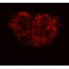
Fig3: Immunohistochemical analysis of paraffin-embedded human tonsil tissue using anti-Thymidine Phosphorylase antibody. The section was pre-treated using heat mediated antigen retrieval with Tris-EDTA buffer (pH 8.0-8.4) for 20 minutes.The tissues were blocked in 5% BSA for 30 minutes at room temperature, washed with ddH2O and PBS, and then probed with the primary antibody for 30 minutes at room temperature. The detection was performed using an HRP conjugated compact polymer system. DAB was used as the chromogen. Tissues were counterstained with hematoxylin and mounted with DPX.
Fig4: Immunohistochemical analysis of paraffin-embedded human liver cancer tissue using anti-Thymidine Phosphorylase antibody. The section was pre-treated using heat mediated antigen retrieval with Tris-EDTA buffer (pH 8.0-8.4) for 20 minutes.The tissues were blocked in 5% BSA for 30 minutes at room temperature, washed with ddH2O and PBS, and then probed with the primary antibody for 30 minutes at room temperature. The detection was performed using an HRP conjugated compact polymer system. DAB was used as the chromogen. Tissues were counterstained with hematoxylin and mounted with DPX.
Fig5: Flow cytometric analysis of Thymidine Phosphorylase was done on Siha cells. The cells were fixed, permeabilized and stained with the primary antibody (red). After incubation of the primary antibody at room temperature for an hour, the cells were stained with a Alexa Fluor 488-conjugated goat anti-rabbit IgG Secondary antibody at 1/500 dilution for 30 minutes.Unlabelled sample was used as a control (cells without incubation with primary antibody; black).
特别提示:本公司的所有产品仅可用于科研实验,严禁用于临床医疗及其他非科研用途!