Anti-Histone Deacetylase 2 antibody
-
概述
- 产品描述In the intact cell, DNA closely associates with histones and other nuclear proteins to form chromatin. The remodeling of chromatin is believed to be a critical component of transcriptional regulation, and a major source of this remodeling is brought about by the acetylation of nucleosomal histones. Acetylation of lysine residues in the amino terminal tail domain of histone results in an allosteric change in the nucleosomal conformation and an increased accessibility to transcription factors by DNA. Conversely, the deacetylation of histones is associated with transcriptional silencing. Several mammalian proteins have been identified as nuclear histone acetylases, including GCN5, PCAF (for p300/CBP-associated factor), p300/CBP and the TFIID subunit TAF II p250. Mammalian HDAC1 (also designated HD1) and HDAC2 (also designated mammalian RPD3), both of which are related to the yeast transcriptional regulator Rpd3p, have been identified as histone deacetylases.
- 产品名称Anti-Histone Deacetylase 2 antibody
- 分子量55 kDa
- 种属反应性Human, Mouse, Rat
- 验证应用WB,ICC,IHC-P,FC
- 抗体类型兔多抗
- 免疫原Recombinant protein within C-terminal human Histone Deacetylase 2.
- 偶联Non-conjugated
-
性能
- 形态Liquid
- 浓度1 mg/mL.
- 存放说明Store at +4℃ after thawing. Aliquot store at -20℃ or -80℃. Avoid repeated freeze / thaw cycles.
- 存储缓冲液1*PBS (pH7.4), 0.2% BSA, 50% Glycerol. Preservative: 0.05% Sodium Azide.
- 亚型IgG
- 纯化方式Protein affinity purified.
- 亚细胞定位Nucleus. Cytoplasm.
- 其它名称
- D10Wsu179e antibody
- HD 2 antibody
- HD2 antibody
more
-
应用
WB: 1:500-1:1,000
ICC: 1:500-1:2,000
IHC-P: 1:100-1:200
FC: 1:50-1:100
-
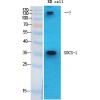
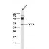
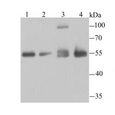
Fig1: Western blot analysis of HDAC2 on different cell lysates using anti-HDAC2 antibody at 1/1,000 dilution.
Positive control:
Lane 1: SH-SY5Y
Lane 2: 293T
Lane 3: Hela
Lane 4: PC-12
Fig2: ICC staining HDAC2 in LOVO cells (green). The nuclear counter stain is DAPI (blue). Cells were fixed in paraformaldehyde, permeabilised with 0.25% Triton X100/PBS.
Fig3: ICC staining HDAC2 in NIH-3T3 cells (green). The nuclear counter stain is DAPI (blue). Cells were fixed in paraformaldehyde, permeabilised with 0.25% Triton X100/PBS.
Fig4: ICC staining HDAC2 in SH-SY5Y cells (green). The nuclear counter stain is DAPI (blue). Cells were fixed in paraformaldehyde, permeabilised with 0.25% Triton X100/PBS.
Fig5: Immunohistochemical analysis of paraffin-embedded human tonsil tissue using anti-HDAC2 antibody. Counter stained with hematoxylin.
Fig6: Immunohistochemical analysis of paraffin-embedded human colon cancer tissue using anti-HDAC2 antibody. Counter stained with hematoxylin.
Fig7: Immunohistochemical analysis of paraffin-embedded human kidney tissue using anti-HDAC2 antibody. Counter stained with hematoxylin.
Fig8: Immunohistochemical analysis of paraffin-embedded mouse brain tissue using anti-HDAC2 antibody. Counter stained with hematoxylin.
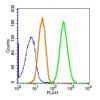
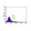
Fig9: Flow cytometric analysis of SH-SY5Y cells with HDAC2 antibody at 1/100 dilution (red) compared with an unlabelled control (cells without incubation with primary antibody; black).
特别提示:本公司的所有产品仅可用于科研实验,严禁用于临床医疗及其他非科研用途!