Anti-DDB1 antibody
-
概述
- 产品描述Damaged DNA binding protein (DDB) is a heterodimer composed of two subunits, p127 and p48, which are designated DDB1 and DDB2, respectively. The DDB heterodimer is involved in repairing DNA damaged by ultraviolet light. Specifically, DDB, also designated UV-damaged DNA binding protein (UV-DDB), xeroderma pigmentosum group E binding factor (XPE-BF) and hepatitis B virus X-associated protein 1 (XAP-1), binds to damaged cyclobutane pyrimidine dimers (CPDs). Mutations in the DDB2 gene are implicated as causes of xeroderma pigmentosum group E, an autosomal recessive disease in which patients are defective in nucleotide excision DNA repair. XPE is characterized by hypersensitivity of the skin to sunlight with a high frequency of skin cancer as well as neurologic abnormalities. The hepatitis B virus (HBV) X protein interacts with DDB1, which may mediate HBx transactivation.
- 产品名称Anti-DDB1 antibody
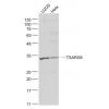
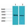
- 分子量127 kDa, additional band 150kDa
- 种属反应性Human,Mouse,Rat
- 验证应用WB,ICC,IHC-P,FC
- 抗体类型兔多抗
- 免疫原Recombinant protein
- 偶联Non-conjugated
-
性能
- 形态Liquid
- 浓度1 mg/mL.
- 存放说明Store at +4℃ after thawing. Aliquot store at -20℃ or -80℃ Avoid repeated freeze / thaw cycles.
- 存储缓冲液1*PBS (pH7.4), 0.2% BSA, 50% Glycerol. Preservative: 0.05% Sodium Azide.
- 亚型IgG
- 纯化方式Protein A purified.
- 亚细胞定位Nucleus. Cytoplasm.
- 其它名称
- Damage specific DNA binding protein 1 antibody
- Damage-specific DNA-binding protein 1 antibody
- DDB 1 antibody
more
-
应用
WB: 1:500-1:1000
ICC: 1:100-1:500
IHC-P: 1:100-1:500
FC: 1:50-1:100
-
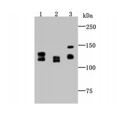
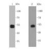
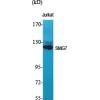
Fig1: Western blot analysis of DDB1 on different cell lysate using anti-DDB1 antibody at 1/1,000 dilution.
Positive control:
Lane 1: Mouse colon tissue
Lane 2: PC-12
Lane 3: Siha
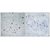
Fig2: ICC staining DDB1 in A549 cells (green). The nuclear counter stain is DAPI (blue). Cells were fixed in paraformaldehyde, permeabilised with 0.25% Triton X100/PBS.
Fig3: ICC staining DDB1 in SH-SY5Y cells (green). The nuclear counter stain is DAPI (blue). Cells were fixed in paraformaldehyde, permeabilised with 0.25% Triton X100/PBS.
Fig4: ICC staining DDB1 in SK-Br-3 cells (green). The nuclear counter stain is DAPI (blue). Cells were fixed in paraformaldehyde, permeabilised with 0.25% Triton X100/PBS.
Fig5: Immunohistochemical analysis of paraffin-embedded rat brain tissue using anti-DDB1 antibody. Counter stained with hematoxylin.
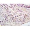
Fig6: Immunohistochemical analysis of paraffin-embedded human breast tissue using anti-DDB1 antibody. Counter stained with hematoxylin.
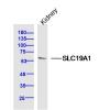
Fig7: Immunohistochemical analysis of paraffin-embedded human kidney tissue using anti-DDB1 antibody. Counter stained with hematoxylin.
Fig8: Flow cytometric analysis of K562 cells with DDB1 antibody at 1/100 dilution (red) compared with an unlabelled control (cells without incubation with primary antibody; black).
特别提示:本公司的所有产品仅可用于科研实验,严禁用于临床医疗及其他非科研用途!