Anti-Paxillin antibody
-
概述
- 产品描述Paxillin is a focal adhesion phosphoprotein that is localized to the cytoskeleton. Phosphorylation of paxillin has been shown to occur in response to PDGF treatment, v-src transformation or cross-linking of integrins. FAK (focal adhesion kinase) and PYK2 have been shown to phosphorylate paxillin. FAK phosphorylates paxillin specifically on Tyr-118 in vitro. However, FAK phosphorylation does not seem to be required for the recruitment of paxillin to cell adhesion sites. Paxillin may play a role in signal transduction, regulation of cell morphology and the recruitment of structural and signaling molecules to focal adhesions. It has been shown that the amount of paxillin is reduced in mitotic cells by proteolytic downregulation and that paxillin is alternatively phosphorylated on serine rather than on tyrosine and serine during mitosis.
- 产品名称Anti-Paxillin antibody
- 分子量68kDa
- 种属反应性Human,Mouse,Rat
- 验证应用WB,IP,IF
- 抗体类型兔多抗
- 免疫原Amino acids 155-268 mapping within an internal region of paxillin of human origin.
- 偶联Non-conjugated
-
性能
- 形态Liquid
- 浓度1 mg/mL.
- 存放说明Store at +4℃
- 存储缓冲液1*TBS (pH7.4), 1%BSA, 40%Glycerol. Preservative: 0.05% Sodium Azide.
- 亚型IgG
- 纯化方式Immunogen affinity purified
- 亚细胞定位Cytoplasm, Cell junction
- 其它名称
- FLJ16691 antibody
- FLJ23042 antibody
- Paired box protein Pax 1 antibody
more
-
应用
WB: 1:100-1:1,000
IP: 1-2 μg per 100-500 μg of total protein (1 ml of cell lysate)
IF: 1:50-1:500
-
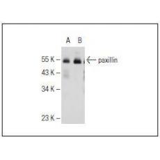
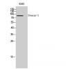
Fig1: Western blot analysis of paxillin expression in non-transfected 293T (A), human paxillin transfected 293T (B) and HISM (C) whole cell lysates.
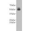
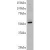
Fig2: Western blot analysis of paxillin expression in CCD-1064Sk (A) and HISM (B) whole cell lysates.
特别提示:本公司的所有产品仅可用于科研实验,严禁用于临床医疗及其他非科研用途!