Anti-SynGAP antibody
-
概述
- 产品描述Synaptic Ras GTPase-activating protein 1, also known as synaptic Ras-GAP 1 or SYNGAP1, is a protein that in humans is encoded by the SYNGAP1 gene. SYNGAP1 is a ras GTPase-activating protein that is critical for the development of cognition and proper synapse function. Mutations in humans can cause intellectual disability or epilepsy. SynGAP1 is a complex protein with several functions that may be regulated temporally via complex isoforms. A well-documented function of SynGAP1 involves NMDA receptor-mediated synaptic plasticity and membrane insertion of AMPA receptors through the suppression of upstream signaling pathways. However, SynGAP1 has also been shown to function cooperatively with Unc51.1 in axon formation. One way SynGAP1 affects these processes is through the MAP kinase signaling pathway by attenuation of Ras signalling. However, alternative splicing and multiple translational start sites have been shown to cause opposing effects, illustrating the importance of multiple functional domains that reside within the c- and n-termini. For example, the expression of an α1 or α2 c-terminal variant of SynGAP1 will either increase or decrease synaptic strength, respectively. Overall, SynGAP1 is essential for development and survival, which is evident as knockout mice die perinatally.
- 产品名称Anti-SynGAP antibody
- 分子量148 kDa
- 种属反应性Human,Mouse,Rat
- 验证应用WB,IHC-P,FC
- 抗体类型兔多抗
- 免疫原Recombinant protein within human SynGAP aa 1100-1250.
- 偶联Non-conjugated
-
性能
- 形态Liquid
- 浓度1 mg/ml.
- 存放说明Store at +4℃ after thawing. Aliquot store at -20℃. Avoid repeated freeze / thaw cycles.
- 存储缓冲液1*PBS (pH7.4), 0.2% BSA, 50% Glycerol. Preservative: 0.05% Sodium Azide.
- 亚型IgG
- 纯化方式Protein affinity purified.
- 亚细胞定位Cytosol.
- 其它名称
- DKFZp761G1421 antibody
- KIAA1938 antibody
- MRD5 antibody
more
-
应用
WB: 1:500-1:2,000
IHC-P: 1:50-1:100
FC:1:50-1:100
-
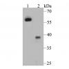
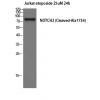
Fig1: Western blot analysis of SynGAP on different lysates. Proteins were transferred to a PVDF membrane and blocked with 5% BSA in PBS for 1 hour at room temperature. The primary antibody was used in 5% BSA at room temperature for 2 hours. Goat Anti-Rabbit IgG - HRP Secondary Antibody (HA1001) at 1:5,000 dilution was used for 1 hour at room temperature.
Positive control:
Lane 1: K562 cell lysate
Lane 2: Mouse brain tissue lysate
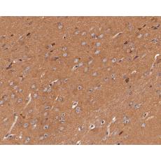
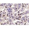
Fig2: Immunohistochemical analysis of paraffin-embedded human terus tissue using anti-SynGAP antibody. The section was pre-treated using heat mediated antigen retrieval with Tris-EDTA buffer (pH 8.0-8.4) for 20 minutes.The tissues were blocked in 5% BSA for 30 minutes at room temperature, washed with ddH2O and PBS, and then probed with the primary antibody for 30 minutes at room temperature. The detection was performed using an HRP conjugated compact polymer system. DAB was used as the chromogen. Tissues were counterstained with hematoxylin and mounted with DPX.
Fig3: Immunohistochemical analysis of paraffin-embedded mouse brain tissue using anti-SynGAP antibody. The section was pre-treated using heat mediated antigen retrieval with Tris-EDTA buffer (pH 8.0-8.4) for 20 minutes.The tissues were blocked in 5% BSA for 30 minutes at room temperature, washed with ddH2O and PBS, and then probed with the primary antibody for 30 minutes at room temperature. The detection was performed using an HRP conjugated compact polymer system. DAB was used as the chromogen. Tissues were counterstained with hematoxylin and mounted with DPX.
Fig4: Flow cytometric analysis of SynGAP was done on SW620 cells. The cells were fixed, permeabilized and stained with the primary antibody (red). After incubation of the primary antibody at room temperature for an hour, the cells were stained with a Alexa Fluor 488-conjugated goat anti-rabbit IgG Secondary antibody at 1/500 dilution for 30 minutes.Unlabelled sample was used as a control (cells without incubation with primary antibody; black).
特别提示:本公司的所有产品仅可用于科研实验,严禁用于临床医疗及其他非科研用途!