Anti-CEACAM6 antibody
-
概述
- 产品描述Cell surface glycoprotein that plays a role in cell adhesion and tumor progression . Intercellular adhesion occurs in a calcium- and fibronectin-independent manner. Mediates homophilic and heterophilic cell adhesion with other carcinoembryonic antigen-related cell adhesion molecules, such as CEACAM5 and CEACAM8 . Heterophilic interaction with CEACAM8 occurs in activated neutrophils . Plays a role in neutrophil adhesion to cytokine-activated endothelial cells . Plays a role as an oncogene by promoting tumor progression; positively regulates cell migration, cell adhesion to endothelial cells and cell invasion . Also involved in the metastatic cascade process by inducing gain resistance to anoikis of pancreatic adenocarcinoma and colorectal carcinoma cells .
- 产品名称Anti-CEACAM6 antibody
- 分子量Predicted band size: 37 kDa.
- 种属反应性Human,Mouse,Rat
- 验证应用WB,IHC-P,ICC,FC
- 抗体类型兔多抗
- 免疫原Recombinant protein corresponding to N terminal Human CEACAM6.
- 偶联Non-conjugated
-
性能
- 形态Liquid
- 浓度1 mg/mL.
- 存放说明Store at +4℃ after thawing. Aliquot store at -20℃. Avoid repeated freeze / thaw cycles.
- 存储缓冲液1*PBS (pH7.4), 0.2% BSA, 50% Glycerol. Preservative: 0.05% Sodium Azide.
- 亚型IgG
- 纯化方式Protein affinity purified.
- 亚细胞定位Cell membrane, Membrane.
- 其它名称
- Carcinoembryonic antigen related cell adhesion molecule 6 antibody
- Carcinoembryonic antigen related cell adhesion molecule 6 (non specific cross reacting antigen) antibody
- Carcinoembryonic antigen-related cell adhesion molecule 6 antibody
more
-
应用
WB: 1:500-1:1,000
IHC-P: 1:50-1:200
ICC: 1:50-1:200
FC: 1:50-1:100
-
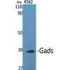
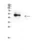
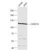
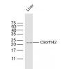
Fig1: Western blot analysis of CEACAM6 on different lysates. Proteins were transferred to a PVDF membrane and blocked with 5% BSA in PBS for 1 hour at room temperature. The primary antibody was used in 5% BSA at room temperature for 2 hours. Goat Anti-Rabbit IgG - HRP Secondary Antibody (HA1001) at 1:5,000 dilution was used for 1 hour at room temperature.
Positive control:
Lane 1: JAR cell lysate
Lane 2: Rat lung tissue lysate
Lane 3: MCF-7 cell lysate
Lane 2: Human lung tissue lysate
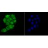
Fig2: ICC staining of CEACAM6 in LOVO cells (green). Formalin fixed cells were permeabilized with 0.1% Triton X-100 in TBS for 10 minutes at room temperature and blocked with 1% Blocker BSA for 15 minutes at room temperature. Cells were probed with the primary antibody for 1 hour at room temperature, washed with PBS. Alexa Fluor®488 Goat anti-Rabbit IgG was used as the secondary antibody at 1/100 dilution. The nuclear counter stain is DAPI (blue).
Fig3: ICC staining of CEACAM6 in MCF-7 cells (green). Formalin fixed cells were permeabilized with 0.1% Triton X-100 in TBS for 10 minutes at room temperature and blocked with 1% Blocker BSA for 15 minutes at room temperature. Cells were probed with the primary antibodyfor 1 hour at room temperature, washed with PBS. Alexa Fluor®488 Goat anti-Rabbit IgG was used as the secondary antibody at 1/100 dilution. The nuclear counter stain is DAPI (blue).
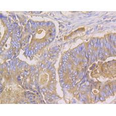
Fig4: Immunohistochemical analysis of paraffin-embedded human lung cancer tissue using anti-CEACAM6 antibody. The section was pre-treated using heat mediated antigen retrieval with Tris-EDTA buffer (pH 8.0-8.4) for 20 minutes.The tissues were blocked in 5% BSA for 30 minutes at room temperature, washed with ddH2O and PBS, and then probed with the primary antibody for 30 minutes at room temperature. The detection was performed using an HRP conjugated compact polymer system. DAB was used as the chromogen. Tissues were counterstained with hematoxylin and mounted with DPX.
Fig5: Immunohistochemical analysis of paraffin-embedded human colon cancer tissue using anti-CEACAM6 antibody. The section was pre-treated using heat mediated antigen retrieval with Tris-EDTA buffer (pH 8.0-8.4) for 20 minutes.The tissues were blocked in 5% BSA for 30 minutes at room temperature, washed with ddH2O and PBS, and then probed with the primary antibody ) for 30 minutes at room temperature. The detection was performed using an HRP conjugated compact polymer system. DAB was used as the chromogen. Tissues were counterstained with hematoxylin and mounted with DPX.
Fig6: Immunohistochemical analysis of paraffin-embedded human appendix cancer tissue using anti-CEACAM6 antibody. The section was pre-treated using heat mediated antigen retrieval with Tris-EDTA buffer (pH 8.0-8.4) for 20 minutes.The tissues were blocked in 5% BSA for 30 minutes at room temperature, washed with ddH2O and PBS, and then probed with the primary antibody for 30 minutes at room temperature. The detection was performed using an HRP conjugated compact polymer system. DAB was used as the chromogen. Tissues were counterstained with hematoxylin and mounted with DPX.
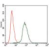
Fig7: Flow cytometric analysis of CEACAM6 was done on MCF-7 cells. The cells were fixed, permeabilized and stained with the primary antibody (red). After incubation of the primary antibody at room temperature for an hour, the cells were stained with a Alexa Fluor 488-conjugated goat anti-rabbit IgG Secondary antibody at 1/500 dilution for 30 minutes.Unlabelled sample was used as a control (cells without incubation with primary antibody; black).
特别提示:本公司的所有产品仅可用于科研实验,严禁用于临床医疗及其他非科研用途!