Anti-GATA3 antibody [A3G6]
-
概述
- 产品描述GATA3 is a transcription factor that in humans is encoded by the GATA3 gene. Studies in animal models and humans indicate that it controls the expression of a wide range of biologically and clinically important genes. The GATA3 transcription factor is critical for the embryonic development of various tissues as well as for inflammatory and humoral immune responses and the proper functioning of the endothelium of blood vessels. GATA3 haploinsufficiency (i.e. lose of one or the two inherited GATA3 genes) results in a congenital disorder termed the Barakat syndrome. Current clinical and laboratory research is focusing on determining the benefits of directly or indirectly blocking the action of GATA3 in inflammatory and allergic diseases such as asthma. It is also proposed to be a clinically important marker for various types of cancer, particularly those of the breast. However, the role, if any, of GATA3 in the development of these cancers is under study and remains unclear.
- 产品名称Anti-GATA3 antibody [A3G6]
- 分子量Predicted band size 48 kDa.
- 种属反应性Human
- 验证应用WB,FC
- 抗体类型小鼠单抗
- 免疫原Synthetic peptide corresponding to N terminal of Human GATA3.
- 偶联Non-conjugated
-
性能
- 形态Liquid
- 浓度2 mg/mL.
- 存放说明Store at +4℃ after thawing. Aliquot store at -20℃. Avoid repeated freeze / thaw cycles.
- 存储缓冲液1*PBS (pH7.4), 0.2% BSA, 50% Glycerol. Preservative: 0.05% Sodium Azide.
- 亚型IgG1
- 纯化方式Protein A purified.
- 亚细胞定位Nucleus.
- 其它名称
- GATA 3 antibody
- GATA binding factor 3 antibody
- GATA binding protein 3 antibody
more
-
应用
WB: 1:1,000-1:5,000
FC: 1:50-1:100
-
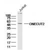
Fig1: Western blot analysis of GATA3 on MCF-7 cell lysate. Proteins were transferred to a PVDF membrane and blocked with 5% BSA in PBS for 1 hour at room temperature. The primary antibody was used in 5% BSA at room temperature for 2 hours. Goat Anti-Mouse IgG - HRP Secondary Antibody (HA1006) at 1:5,000 dilution was used for 1 hour at room temperature.
Specific bands were detected for GATA3 full length (FL) at approximately 52 kDa and the splice form (SF) at approximately 39 kDa (as indicated).
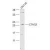
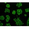
Fig2: Flow cytometric analysis of GATA3 was done on jurkat cells. The cells were fixed, permeabilized and stained with the primary antibody (, 1/100) (red). After incubation of the primary antibody at room temperature for an hour, the cells were stained with a Alexa Fluor 488-conjugated goat anti-Mouse IgG Secondary antibody at 1/500 dilution for 30 minutes.Unlabelled sample was used as a control (cells without incubation with primary antibody; black).
特别提示:本公司的所有产品仅可用于科研实验,严禁用于临床医疗及其他非科研用途!

![Anti-GATA3 antibody [A3G6]](images/202012/goods_img/92960_G_1608462629004.jpg)