Anti-SDF1 antibody
-
概述
- 产品描述Chemoattractant active on T-lymphocytes and monocytes but not neutrophils. Activates the C-X-C chemokine receptor CXCR4 to induce a rapid and transient rise in the level of intracellular calcium ions and chemotaxis. SDF-1-beta(3-72) and SDF-1-alpha(3-67) show a reduced chemotactic activity. Binding to cell surface proteoglycans seems to inhibit formation of SDF-1-alpha(3-67) and thus to preserve activity on local sites. Also binds to atypical chemokine receptor ACKR3, which activates the beta-arrestin pathway and acts as a scavenger receptor for SDF-1. Binds to the allosteric site (site 2) of integrins and activates integrins ITGAV:ITGB3, ITGA4:ITGB1 and ITGA5:ITGB1 in a CXCR4-independent manner . Acts as a positive regulator of monocyte migration and a negative regulator of monocyte adhesion via the LYN kinase. Stimulates migration of monocytes and T-lymphocytes through its receptors, CXCR4 and ACKR3, and decreases monocyte adherence to surfaces coated with ICAM-1, a ligand for beta-2 integrins. SDF1A/CXCR4 signaling axis inhibits beta-2 integrin LFA-1 mediated adhesion of monocytes to ICAM-1 through LYN kinase. Inhibits CXCR4-mediated infection by T-cell line-adapted HIV-1. Plays a protective role after myocardial infarction. Induces down-regulation and internalization of ACKR3 expressed in various cells. Has several critical functions during embryonic development; required for B-cell lymphopoiesis, myelopoiesis in bone marrow and heart ventricular septum formation.
- 产品名称Anti-SDF1 antibody
- 分子量Predicted band size 10 kDa.
- 种属反应性Human,Mouse,Rat
- 验证应用WB,IHC-P,ICC,FC
- 抗体类型兔多抗
- 免疫原Recombinant protein within Huamn SDF1 alpha aa 1-93.
- 偶联Non-conjugated
-
性能
- 形态Liquid
- 浓度1 mg/ml.
- 存放说明Store at +4℃ after thawing. Aliquot store at -20℃. Avoid repeated freeze / thaw cycles.
- 存储缓冲液1*PBS (pH7.4), 0.2% BSA, 50% Glycerol. Preservative: 0.05% Sodium Azide.
- 亚型IgG
- 纯化方式Protein affinity purified.
- 亚细胞定位Secreted.
- 其它名称
- 12-O-tetradecanoylphorbol 13-acetate repressed protein 1 antibody
- AI174028 antibody
- C-X-C motif chemokine 12 antibody
more
-
应用
WB: 1:500
IHC-P: 1:50-1:200
ICC: 1:100-1:500
FC: 1:50-1:100
-
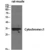
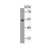
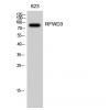
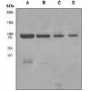
Fig1: Western blot analysis of SDF1 on recombinant protein lysate. Proteins were transferred to a PVDF membrane and blocked with 5% BSA in PBS for 1 hour at room temperature. The primary antibody was used in 5% BSA at room temperature for 2 hours. Goat Anti-Rabbit IgG - HRP Secondary Antibody (HA1001) at 1:5,000 dilution was used for 1 hour at room temperature.
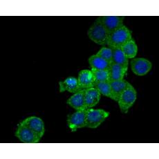
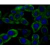
Fig2: ICC staining of SDF1 alpha in 293T cells (green). Formalin fixed cells were permeabilized with 0.1% Triton X-100 in TBS for 10 minutes at room temperature and blocked with 1% Blocker BSA for 15 minutes at room temperature. Cells were probed with the primary antibody for 1 hour at room temperature, washed with PBS. Alexa Fluor®488 Goat anti-Rabbit IgG was used as the secondary antibody at 1/100 dilution. The nuclear counter stain is DAPI (blue).
Fig3: ICC staining of SDF1 alpha in LOVO cells (green). Formalin fixed cells were permeabilized with 0.1% Triton X-100 in TBS for 10 minutes at room temperature and blocked with 1% Blocker BSA for 15 minutes at room temperature. Cells were probed with the primary antibody for 1 hour at room temperature, washed with PBS. Alexa Fluor®488 Goat anti-Rabbit IgG was used as the secondary antibody at 1/100 dilution. The nuclear counter stain is DAPI (blue).
Fig4: Immunohistochemical analysis of paraffin-embedded Rat kidney tissue using anti-SDF1 alpha antibody. The section was pre-treated using heat mediated antigen retrieval with Tris-EDTA buffer (pH 8.0-8.4) for 20 minutes.The tissues were blocked in 5% BSA for 30 minutes at room temperature, washed with ddH2O and PBS, and then probed with the primary antibody for 30 minutes at room temperature. The detection was performed using an HRP conjugated compact polymer system. DAB was used as the chromogen. Tissues were counterstained with hematoxylin and mounted with DPX.
Fig5: Immunohistochemical analysis of paraffin-embedded Human colon cancer tissue using anti-SDF1 alpha antibody. The section was pre-treated using heat mediated antigen retrieval with Tris-EDTA buffer (pH 8.0-8.4) for 20 minutes.The tissues were blocked in 5% BSA for 30 minutes at room temperature, washed with ddH2O and PBS, and then probed with the primary antibody for 30 minutes at room temperature. The detection was performed using an HRP conjugated compact polymer system. DAB was used as the chromogen. Tissues were counterstained with hematoxylin and mounted with DPX.
Fig6: Immunohistochemical analysis of paraffin-embedded Human kidney tissue using anti-SDF1 alpha antibody. The section was pre-treated using heat mediated antigen retrieval with Tris-EDTA buffer (pH 8.0-8.4) for 20 minutes.The tissues were blocked in 5% BSA for 30 minutes at room temperature, washed with ddH2O and PBS, and then probed with the primary antibody for 30 minutes at room temperature. The detection was performed using an HRP conjugated compact polymer system. DAB was used as the chromogen. Tissues were counterstained with hematoxylin and mounted with DPX.
Fig7: Immunohistochemical analysis of paraffin-embedded mouse colon tissue using anti-SDF1 alpha antibody. The section was pre-treated using heat mediated antigen retrieval with Tris-EDTA buffer (pH 8.0-8.4) for 20 minutes.The tissues were blocked in 5% BSA for 30 minutes at room temperature, washed with ddH2O and PBS, and then probed with the primary antibody for 30 minutes at room temperature. The detection was performed using an HRP conjugated compact polymer system. DAB was used as the chromogen. Tissues were counterstained with hematoxylin and mounted with DPX.
Fig8: Flow cytometric analysis of SDF1 alpha was done on HepG2 cells. The cells were fixed, permeabilized and stained with the primary antibody (red). After incubation of the primary antibody at room temperature for an hour, the cells were stained with a Alexa Fluor 488-conjugated goat anti-rabbit IgG Secondary antibody at 1/500 dilution for 30 minutes.Unlabelled sample was used as a control (cells without incubation with primary antibody; black).
特别提示:本公司的所有产品仅可用于科研实验,严禁用于临床医疗及其他非科研用途!