Anti-SLC8B1 antibody
-
概述
- 产品描述Mitochondrial sodium/calcium antiporter that mediates sodium-dependent calcium efflux from mitochondrion, by mediating the exchange of 3 sodium ions per 1 calcium ion . Plays a central role in mitochondrial calcium homeostasis by mediating mitochondrial calcium extrusion: calcium efflux is essential for mitochondrial function and cell survival, notably in cardiomyocytes (By similarity). Regulates rates of glucose-dependent insulin secretion in pancreatic beta-cells during the first phase of insulin secretion: acts by mediating efflux of calcium from mitochondrion, thereby affecting cytoplasmic calcium responses . Required for store-operated Ca2+ entry (SOCE) and Ca2+ release-activated Ca2+ (CRAC) channel regulation: sodium transport by SLC8B1 leads to promote calcium-shuttling that modulates mitochondrial redox status, thereby regulating SOCE activity . Involved in B-lymphocyte chemotaxis (By similarity). Able to transport Ca2+ in exchange of either Li+ or Na+, explaining how Li+ catalyzes Ca2+ exchange . In contrast to other members of the family its function is independent of K+ .
- 产品名称Anti-SLC8B1 antibody
- 分子量64 kDa
- 种属反应性Human,Mouse,Rat
- 验证应用WB,IHC-P,ICC,FC
- 抗体类型兔多抗
- 免疫原Recombinant protein within Human SLC8B1 aa 200-400.
- 偶联Non-conjugated
-
性能
- 形态Liquid
- 浓度1 mg/ml.
- 存放说明Store at +4℃ after thawing. Aliquot store at -20℃. Avoid repeated freeze / thaw cycles.
- 存储缓冲液1*PBS (pH7.4), 0.2% BSA, 50% Glycerol. Preservative: 0.05% Sodium Azide.
- 亚型IgG
- 纯化方式Protein affinity purified.
- 亚细胞定位Membrane, Mitochondrion, Mitochondrion inner membrane.
- 其它名称
- FLJ22233 antibody
- Na(+)/K(+)/Ca(2+) exchange protein 6 antibody
- Na(+)/K(+)/Ca(2+)-exchange protein 6 antibody
more
-
应用
WB: 1:500-1:1000
IHC-P: 1:50-1:200
ICC: 1:50-1:200
FC: 1:50-1:100
-
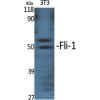
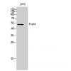
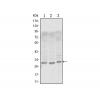
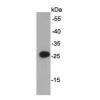
Fig1: Western blot analysis of SLC8B1 on mouse tonsil tissue lysate. Proteins were transferred to a PVDF membrane and blocked with 5% BSA in PBS for 1 hour at room temperature. The primary antibody ( was used in 5% BSA at room temperature for 2 hours. Goat Anti-Rabbit IgG - HRP Secondary Antibody (HA1001) at 1:5,000 dilution was used for 1 hour at room temperature.
Fig2: ICC staining of SLC8B1 in SHSY5Y cells (green). Formalin fixed cells were permeabilized with 0.1% Triton X-100 in TBS for 10 minutes at room temperature and blocked with 1% Blocker BSA for 15 minutes at room temperature. Cells were probed with the primary antibody for 1 hour at room temperature, washed with PBS. Alexa Fluor®488 Goat anti-Rabbit IgG was used as the secondary antibody at 1/100 dilution. The nuclear counter stain is DAPI (blue).
Fig3: ICC staining of SLC8B1 in SKOV-3 cells (green). Formalin fixed cells were permeabilized with 0.1% Triton X-100 in TBS for 10 minutes at room temperature and blocked with 1% Blocker BSA for 15 minutes at room temperature. Cells were probed with the primary antibody for 1 hour at room temperature, washed with PBS. Alexa Fluor®488 Goat anti-Rabbit IgG was used as the secondary antibody at 1/100 dilution. The nuclear counter stain is DAPI (blue).
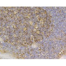
Fig4: Immunohistochemical analysis of paraffin-embedded rat kidney tissue using anti-SLC8B1 antibody. The section was pre-treated using heat mediated antigen retrieval with Tris-EDTA buffer (pH 8.0-8.4) for 20 minutes.The tissues were blocked in 5% BSA for 30 minutes at room temperature, washed with ddH2O and PBS, and then probed with the primary antibody for 30 minutes at room temperature. The detection was performed using an HRP conjugated compact polymer system. DAB was used as the chromogen. Tissues were counterstained with hematoxylin and mounted with DPX.
Fig5: Immunohistochemical analysis of paraffin-embedded human tonsil tissue using anti-SLC8B1 antibody. The section was pre-treated using heat mediated antigen retrieval with Tris-EDTA buffer (pH 8.0-8.4) for 20 minutes.The tissues were blocked in 5% BSA for 30 minutes at room temperature, washed with ddH2O and PBS, and then probed with the primary antibody ( for 30 minutes at room temperature. The detection was performed using an HRP conjugated compact polymer system. DAB was used as the chromogen. Tissues were counterstained with hematoxylin and mounted with DPX.
Fig6: Immunohistochemical analysis of paraffin-embedded human pancreas tissue using anti-SLC8B1 antibody. The section was pre-treated using heat mediated antigen retrieval with Tris-EDTA buffer (pH 8.0-8.4) for 20 minutes.The tissues were blocked in 5% BSA for 30 minutes at room temperature, washed with ddH2O and PBS, and then probed with the primary antibody for 30 minutes at room temperature. The detection was performed using an HRP conjugated compact polymer system. DAB was used as the chromogen. Tissues were counterstained with hematoxylin and mounted with DPX.
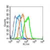
Fig7: Flow cytometric analysis of SLC8B1 was done on Siha cells. The cells were fixed, permeabilized and stained with the primary antibody (red). After incubation of the primary antibody at room temperature for an hour, the cells were stained with a Alexa Fluor 488-conjugated goat anti-rabbit IgG Secondary antibody at 1/500 dilution for 30 minutes.Unlabelled sample was used as a control (cells without incubation with primary antibody; black).
特别提示:本公司的所有产品仅可用于科研实验,严禁用于临床医疗及其他非科研用途!