Anti-Beta Actin antibody
-
概述
- 产品描述Beta-actin (human gene and protein abbreviation ACTB/ACTB) is one of six different actin isoforms which have been identified in humans. This is one of the two nonmuscle cytoskeletal actins. Actins are highly conserved proteins that are involved in cell motility, structure and integrity. Alpha actins are a major constituent of the contractile apparatus. Beta-actin has been shown to interact with SPTBN2. In addition, RNA-binding protein Sam68 was found to interact with the mRNA encoding β-actin, which regulates the synaptic formation of the dendritic spines with its cytoskeletal components. Beta-actin has been shown to activate eNOS, thereby increasing NO production. An eight-amino acid residue (326-333) in actin has been shown to mediate the interaction between actin and eNOS. Recurrent mutations in this gene have been associated to cases of diffuse large B-cell lymphoma. Beta actin is usually used as a loading control, for among others, the integrity of cells, protein degradation, in PCR and Western blotting. Its molecular weight is approximately 42 kDa.
- 产品名称Anti-Beta Actin antibody
- 分子量42 kDa
- 种属反应性Human,Mouse,Rat
- 验证应用WB,ICC,IHC-P,FC
- 抗体类型兔多抗
- 免疫原Synthetic peptide within N-terminal residues of Human beta actin.
- 偶联Non-conjugated
-
性能
- 形态Liquid
- 浓度1mg/ml
- 存放说明Store at +4℃ after thawing. Aliquot store at -20℃. Avoid repeated freeze / thaw cycles.
- 存储缓冲液1*PBS (pH7.4), 0.2% BSA, 50% Glycerol. Preservative: 0.05% Sodium Azide.
- 亚型IgG
- 纯化方式Peptide affinity purified.
- 亚细胞定位Cytoskeleton, Nucleus.
- 其它名称
- A26C1A antibody
- A26C1B antibody
- ACTB antibody
more
-
应用
WB: 1:1,000-2,000
ICC: 1:50-1:200
IHC-P: 1:50-1:200
FC: 1:50-1:200
-
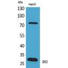
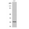
Fig1: Western blot analysis of β-Actin on different cell lysate. Proteins were transferred to a PVDF membrane and blocked with 5% BSA in PBS for 1 hour at room temperature. The primary antibody was used at a 1:50,000 dilution in 5% BSA at room temperature for 2 hours. Goat Anti-rabbit IgG - HRP Secondary Antibody (HA1001) at 1:5,000 dilution was used for 1 hour at room temperature.
Positive control:
Lane A: PC12 cell lysates
Lane B: Hela cell lysates
Lane C: NIH/3T3 cell lysates
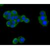
Fig2: ICC staining of β-Actin in MCF-7 cells (green). Formalin fixed cells were permeabilized with 0.1% Triton X-100 in TBS for 10 minutes at room temperature and blocked with 1% Blocker BSA for 15 minutes at room temperature. Cells were probed with the primary antibody for 1 hour at room temperature, washed with PBS. Alexa Fluor®488 Goat anti-Rabbit IgG was used as the secondary antibody at 1/100 dilution. The nuclear counter stain is DAPI (blue).
Fig3: Immunohistochemical analysis of paraffin-embedded human kidney tissue using anti-β-Actin antibody. The section was pre-treated using heat mediated antigen retrieval with sodium citrate buffer (pH 6.0) for 20 minutes. The tissues were blocked in 5% BSA for 30 minutes at room temperature, washed with ddH2O and PBS, and then probed with the primary antibody for 30 minutes at room temperature. The detection was performed using an HRP conjugated compact polymer system. DAB was used as the chromogen. Tissues were counterstained with hematoxylin and mounted with DPX.
Fig4: Immunohistochemical analysis of paraffin-embedded human tonsil tissue using anti-β-Actin antibody. The section was pre-treated using heat mediated antigen retrieval with sodium citrate buffer (pH 6.0) for 20 minutes. The tissues were blocked in 5% BSA for 30 minutes at room temperature, washed with ddH2O and PBS, and then probed with the primary antibody for 30 minutes at room temperature. The detection was performed using an HRP conjugated compact polymer system. DAB was used as the chromogen. Tissues were counterstained with hematoxylin and mounted with DPX.
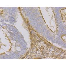
Fig5: Immunohistochemical analysis of paraffin-embedded human colon cancer tissue using anti-β-Actin antibody. The section was pre-treated using heat mediated antigen retrieval with sodium citrate buffer (pH 6.0) for 20 minutes. The tissues were blocked in 5% BSA for 30 minutes at room temperature, washed with ddH2O and PBS, and then probed with the primary antibody for 30 minutes at room temperature. The detection was performed using an HRP conjugated compact polymer system. DAB was used as the chromogen. Tissues were counterstained with hematoxylin and mounted with DPX.
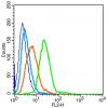
Fig6: Flow cytometric analysis of β-Actin was done on HT-29 cells. The cells were fixed, permeabilized and stained with the primary antibody (red). After incubation of the primary antibody at room temperature for an hour, the cells were stained with a Alexa Fluor 488-conjugated goat anti-rabbit IgG Secondary antibody at 1/500 dilution for 30 minutes.Unlabelled sample was used as a control (cells without incubation with primary antibody; black).
特别提示:本公司的所有产品仅可用于科研实验,严禁用于临床医疗及其他非科研用途!