Anti-USP21 antibody
-
概述
- 产品描述The ubiquitin (Ub) pathway involves three sequential enzymatic steps that facilitate the conjugation of Ub and Ub-like molecules to specific protein substrates. Through the use of a wide range of enzymes that can add or remove ubiquitin, the Ub pathway controls many intracellular processes such as signal transduction, transcriptional activation and cell cycle progression. USP21 (ubiquitin specific peptidase 21), also known as USP16 or USP23, is a 565 amino acid protein that belongs to the C19 peptidase family of ubiquitin carboxy-terminal hydrolases. Capable of removing ubiquitin from ubiquitinated proteins, USP21 plays a role in signal transduction and can also remove NEDD8 from NEDD8-conjugated proteins, possibly functioning to influence NEDD8-mediated protein proteolysis. Multiple isoforms of USP21 exist due to alternative splicing events.
- 产品名称Anti-USP21 antibody
- 分子量63 kDa
- 种属反应性Human,Mouse,Rat
- 验证应用WB,IHC-P,FC
- 抗体类型兔多抗
- 免疫原Recombinant protein within Human USP21 aa 300-450.
- 偶联Non-conjugated
-
性能
- 形态Liquid
- 浓度1 mg/ml.
- 存放说明Store at +4℃ after thawing. Aliquot store at -20℃. Avoid repeated freeze / thaw cycles.
- 存储缓冲液1*PBS (pH7.4), 0.2% BSA, 50% Glycerol. Preservative: 0.05% Sodium Azide.
- 亚型IgG
- 纯化方式Protein affinity purified.
- 亚细胞定位Cytoplasm. Nucleus.
- 其它名称
- Deubiquitinating enzyme 21 antibody
- deubiquitinating enzyme 23 antibody
- EC 3.1.2.15 antibody
more
-
应用
WB: 1:500-1:2000
IHC-P: 1:50-1;200
FC: 1:50-1;200
-
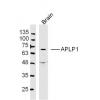
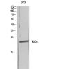
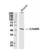
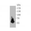
Fig1: Western blot analysis of USP21 on SHSY5Y cell lysates. Proteins were transferred to a PVDF membrane and blocked with 5% BSA in PBS for 1 hour at room temperature. The primary antibody was used in 5% BSA at room temperature for 2 hours. Goat Anti-Rabbit IgG - HRP Secondary Antibody (HA1001) at 1:5,000 dilution was used for 1 hour at room temperature.
Fig2: Immunohistochemical analysis of paraffin-embedded Rat kidney tissue using anti-USP21 antibody. The section was pre-treated using heat mediated antigen retrieval with Tris-EDTA buffer (pH 8.0-8.4) for 20 minutes.The tissues were blocked in 5% BSA for 30 minutes at room temperature, washed with ddH2O and PBS, and then probed with the primary antibody for 30 minutes at room temperature. The detection was performed using an HRP conjugated compact polymer system. DAB was used as the chromogen. Tissues were counterstained with hematoxylin and mounted with DPX.
Fig3: Immunohistochemical analysis of paraffin-embedded human fetal skeletal muscle tissue using anti-USP21 antibody. The section was pre-treated using heat mediated antigen retrieval with Tris-EDTA buffer (pH 8.0-8.4) for 20 minutes.The tissues were blocked in 5% BSA for 30 minutes at room temperature, washed with ddH2O and PBS, and then probed with the primary antibody for 30 minutes at room temperature. The detection was performed using an HRP conjugated compact polymer system. DAB was used as the chromogen. Tissues were counterstained with hematoxylin and mounted with DPX.
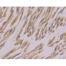
Fig4: Immunohistochemical analysis of paraffin-embedded Mouse heart tissue using anti-USP21 antibody. The section was pre-treated using heat mediated antigen retrieval with Tris-EDTA buffer (pH 8.0-8.4) for 20 minutes.The tissues were blocked in 5% BSA for 30 minutes at room temperature, washed with ddH2O and PBS, and then probed with the primary antibody for 30 minutes at room temperature. The detection was performed using an HRP conjugated compact polymer system. DAB was used as the chromogen. Tissues were counterstained with hematoxylin and mounted with DPX.
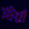
Fig5: Flow cytometric analysis of USP21 was done on SHSY5Y cells. The cells were fixed, permeabilized and stained with the primary antibody (red). After incubation of the primary antibody at room temperature for an hour, the cells were stained with a Alexa Fluor 488-conjugated goat anti-rabbit IgG Secondary antibody at 1/500 dilution for 30 minutes.Unlabelled sample was used as a control (cells without incubation with primary antibody; green).
特别提示:本公司的所有产品仅可用于科研实验,严禁用于临床医疗及其他非科研用途!