Anti-RanBP9 antibody
-
概述
- 产品描述Functions in nuclear protein import as nuclear transport receptor. Serves as receptor for nuclear localization signals (NLS) in cargo substrates. Is thought to mediate docking of the importin/substrate complex to the nuclear pore complex (NPC) through binding to nucleoporin and the complex is subsequently translocated through the pore by an energy requiring, Ran-dependent mechanism. At the nucleoplasmic side of the NPC, Ran binds to the importin, the importin/substrate complex dissociates and importin is re-exported from the nucleus to the cytoplasm where GTP hydrolysis releases Ran. The directionality of nuclear import is thought to be conferred by an asymmetric distribution of the GTP- and GDP-bound forms of Ran between the cytoplasm and nucleus. Mediates the nuclear import of RPS7, RPL18A, RPL6, histone H2A, histone H2B and histone. Prevents the cytoplasmic aggregation of RPS7 and RPL18A by shielding exposed basic domains. Mediates the nuclear import of actin .
- 产品名称Anti-RanBP9 antibody
- 分子量116 kDa
- 种属反应性Human,Mouse,Rat
- 验证应用WB,FC
- 抗体类型兔多抗
- 免疫原Recombinant protein within human RanBP9 aa 800-1041.
-
性能
- 形态Liquid
- 浓度1 mg/mL.
- 存放说明Store at +4℃ after thawing. Aliquot store at -20℃. Avoid repeated freeze / thaw cycles.
- 存储缓冲液1*TBS (pH7.4), 0.2% BSA, 50% Glycerol. Preservative: 0.05% Sodium Azide.
- 亚型IgG
- 纯化方式Protein affinity purified.
- 亚细胞定位Cytoplasm, Nucleus.
- 其它名称
- Imp9 antibody
- Imp9a antibody
- Imp9b antibody
more
-
应用
WB: 1:500-1:2,000
FC: 1:50-1:100
-
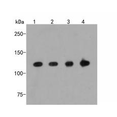
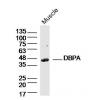
Fig1: Western blot analysis of RanBP9 on different lysates. Proteins were transferred to a PVDF membrane and blocked with 5% BSA in PBS for 1 hour at room temperature. The primary antibody was used in 5% BSA at room temperature for 2 hours. Goat Anti-Rabbit IgG - HRP Secondary Antibody (HA1001) at 1:5,000 dilution was used for 1 hour at room temperature.
Positive control:
Lane 1: SKBR-3 cell lysate
Lane 2: 293 cell lysate
Lane 3: Mouse lung tissue lysate
Lane 4: Rat brain tissue lysate
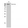
Fig2: Flow cytometric analysis of RanBP9 was done on F9 cells. The cells were fixed, permeabilized and stained with the primary antibody (red). After incubation of the primary antibody at room temperature for an hour, the cells were stained with a Alexa Fluor 488-conjugated Goat anti-Rabbit IgG Secondary antibody at 1/1000 dilution for 30 minutes.Unlabelled sample was used as a control (cells without incubation with primary antibody; black).
特别提示:本公司的所有产品仅可用于科研实验,严禁用于临床医疗及其他非科研用途!