Anti-CD133 antibody
-
概述
- 产品描述CD133 antigen, also known as prominin-1, is a glycoprotein that in humans is encoded by the PROM1 gene. It is a member of pentaspan transmembrane glycoproteins, which specifically localize to cellular protrusions. When embedded in the cell membrane, the membrane topology of prominin-1 is such that the N-terminus extends into the extracellular space and the C-terminus resides in the intracellular compartment. The protein consists of five transmembrane segments, with the first and second segments and the third and fourth segments connected by intracellular loops while the second and third as well as fourth and fifth transmembrane segments are connected by extracellular loops. While the precise function of CD133 remains unknown, it has been proposed that it acts as an organizer of cell membrane topology. CD133 is expressed in hematopoietic stem cells, endothelial progenitor cells, glioblastoma, neuronal and glial stem cells, various pediatric brain tumors, as well as adult kidney, mammary glands, trachea, salivary glands, uterus, placenta, digestive tract, testes, and some other cell types.
- 产品名称Anti-CD133 antibody
- 分子量Predicted band size 97 kDa.
- 种属反应性Human,Mouse,Rat
- 验证应用WB,FC
- 抗体类型兔多抗
- 免疫原Synthetic peptide within human CD133 aa 800-865.
-
性能
- 形态Liquid
- 浓度1 mg/mL.
- 存放说明Store at +4℃ after thawing. Aliquot store at -20℃. Avoid repeated freeze / thaw cycles.
- 存储缓冲液1*TBS (pH7.4), 0.2% BSA, 50% Glycerol. Preservative: 0.05% Sodium Azide.
- 亚型IgG
- 纯化方式Peptide affinity purified.
- 亚细胞定位Cell membrane.
- 其它名称
- AC133 antibody
- Antigen AC133 antibody
- CD133 antibody
more
-
应用
WB: 1:500
FC: 1:50-1:100
-
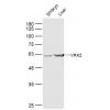
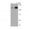
Fig1: Western blot analysis of CD133 on human stomach tissue lysates. Proteins were transferred to a PVDF membrane and blocked with 5% BSA in PBS for 1 hour at room temperature. The primary antibody was used in 5% BSA at room temperature for 2 hours. Goat Anti-Rabbit IgG - HRP Secondary Antibody (HA1001) at 1:5,000 dilution was used for 1 hour at room temperature.
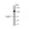
Fig2: Western blot analysis of CD133 on PC-12 cell lysates. Proteins were transferred to a PVDF membrane and blocked with 5% BSA in PBS for 1 hour at room temperature. The primary antibody ( was used in 5% BSA at room temperature for 2 hours. Goat Anti-Rabbit IgG - HRP Secondary Antibody (HA1001) at 1:5,000 dilution was used for 1 hour at room temperature.
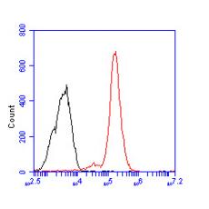
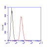
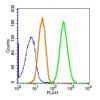
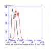
Fig3: Flow cytometric analysis of CD133 was done on F9 cells. The cells were fixed, permeabilized and stained with the primary antibody (red). After incubation of the primary antibody at room temperature for an hour, the cells were stained with a Alexa Fluor 488-conjugated Goat anti-Rabbit IgG Secondary antibody at 1/1000 dilution for 30 minutes.Unlabelled sample was used as a control (cells without incubation with primary antibody; black).
特别提示:本公司的所有产品仅可用于科研实验,严禁用于临床医疗及其他非科研用途!