Anti-β-tubulin antibody
-
概述
- 产品描述Tubulin is one of several members of a small family of globular proteins. The most common members of the tubulin family are α-tubulin and β-tubulin. The beta-tubulin (relative molecular weight about 50 kDa) is counterpart of alpha-tubulin in tubulin heterodimer, it is coded by multiple tubulin genes and it is also posttranslationally modified. Heterogeneity of subunit is concentrated in C-terminal structural domain. Beta-Tubulin may have bound GTP or GDP. Under certain conditions β-tubulin can hydrolyze its bound GTP to GDP plus Pi, release the Pi, and exchange the GDP for GTP.
- 产品名称Anti-β-tubulin antibody
- 分子量50 kDa
- 种属反应性Human,Mouse,Rat,Zebrafish
- 验证应用WB, ICC, IHC-P, FC
- 抗体类型小鼠单抗
- 免疫原peptide
- 偶联Non-conjugated
-
性能
- 形态Liquid
- 浓度2 mg/mL.
- 存放说明Store at +4℃ after thawing. Aliquot store at -20℃ or -80℃. Avoid repeated freeze / thaw cycles.
- 存储缓冲液1*PBS (pH7.4), 0.2% BSA, 40% Glycerol. Preservative: 0.05% Sodium Azide.
- 亚型IgG1
- 纯化方式Protein A purified.
- 亚细胞定位Cytoplasm ,cytoskeleton
- 其它名称
- Beta 4 tubulin antibody
- Beta 5 tubulin antibody
- BetaTubulin antibody
more
-
应用
WB: 1:5,000-1:10,000
ICC: 1:200
IHC-P: 1:20
FC: 1:50-1:100
-
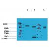
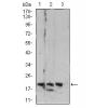
Fig1: Western blot analysis of β-tubulin on different cell lysates using anti-β-tubulin antibody at 1/5000 dilution.
Positive control:
Lane 1: NCCIT
Lane 2: NIH/3T3
Lane 3: PC12
Lane 4: Mouse heart
Lane 5: F9
Lane 6: zebrafish brain
Lane 7: Hela
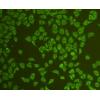
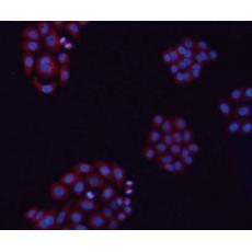
Fig2: ICC staining β-tubulin in Hela cells (red). The nuclear counter stain is DAPI (blue). Cells were fixed in paraformaldehyde, permeabilised with 0.25% Triton X100/PBS.
Fig3: ICC stainingβ-tubulin in HepG2 cells (red). The nuclear counter stain is DAPI (blue). Cells were fixed in paraformaldehyde, permeabilised with 0.25% Triton X100/PBS.
Fig4: ICC stainingβ-tubulin in NIH/3T3 cells (red). The nuclear counter stain is DAPI (blue). Cells were fixed in paraformaldehyde, permeabilised with 0.25% Triton X100/PBS.
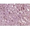
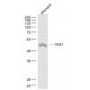
Fig5: Immunohistochemical analysis of paraffin-embedded mouse brain tissue using anti-β-tubulin antibody. Counter stained with hematoxylin.
Fig6: Flow cytometric analysis of HeLa cells with β-tubulin antibody at 1/50 dilution (blue) compared with an unlabelled control (cells without incubation with primary antibody; red). Goat anti mouse IgG (FITC) was used as the secondary antibody.
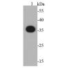
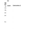
Fig7: Western blot analysis of β-tubulin on hybrid fish (crucian-carp) brain tissue lysate using anti-β-tubulin antibody at 1/500 dilution.
特别提示:本公司的所有产品仅可用于科研实验,严禁用于临床医疗及其他非科研用途!