Anti-AKT1 antibody
-
概述
- 产品描述The serine-threonine protein kinase AKT1 is catalytically inactive in serum-starved primary and immortalized fibroblasts. AKT1 and the related AKT2 are activated by platelet-derived growth factor. The activation is rapid and specific, and it is abrogated by mutations in the pleckstrin homology domain of AKT1. It was shown that the activation occurs through phosphatidylinositol 3-kinase. In the developing nervous system AKT is a critical mediator of growth factor-induced neuronal survival. Survival factors can suppress apoptosis in a transcription-independent manner by activating the serine/threonine kinase AKT1, which then phosphorylates and inactivates components of the apoptotic machinery. Mice lacking Akt1 display a 25% reduction in body mass, indicating that Akt1 is critical for transmitting growth-promoting signals, most likely via the igf1 receptor. Mice lacking Akt1 are also resistant to cancer: They experience considerable delay in tumor growth initiated by the large T antigen or the Neuoncogene.
- 产品名称Anti-AKT1 antibody
- 分子量56 kDa
- 种属反应性Human,Mouse
- 验证应用WB,ICC,IHC-P,FC
- 抗体类型小鼠单抗
- 免疫原peptide
- 偶联Non-conjugated
-
性能
- 形态Liquid
- 浓度2 mg/mL.
- 存放说明Store at +4℃ after thawing. Aliquot store at -20℃ or -80℃. Avoid repeated freeze / thaw cycles.
- 存储缓冲液1*PBS (pH7.4), 0.2% BSA, 40% Glycerol. Preservative: 0.05% Sodium Azide.
- 亚型IgG2b
- 纯化方式Protein A purified.
- 亚细胞定位Nucleus, cytoplasm, cell membrane.
- 其它名称
- AKT 1 antibody
- AKT antibody
- AKT1 antibody
more
-
应用
WB: 1:2,000
IHC-P: 1:100-1:200
ICC: 1:100-1:200
FC: 100-1:200
-
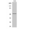
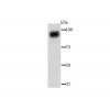
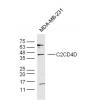
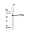
Fig1: Western blot analysis of Akt1 on different cell lysates using anti-Akt1 antibody at 1/2000 dilution.
Positive control:
Lane 1: HepG2
Lane 2: MCF-7
Lane 3: Hela
Fig2: ICC staining Akt1 in Hela cells (red). Cells were fixed in paraformaldehyde, permeabilised with 0.25% Triton X100/PBS.
Fig3: ICC staining Akt1 in HepG2 cells (red). Cells were fixed in paraformaldehyde, permeabilised with 0.25% Triton X100/PBS.
Fig4: ICC staining Akt1 in MCF-7 cells (red). Cells were fixed in paraformaldehyde, permeabilised with 0.25% Triton X100/PBS.
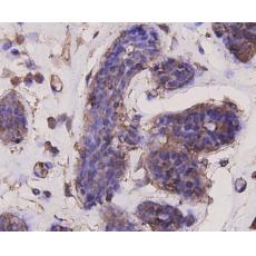
Fig5: Immunohistochemical analysis of paraffin-embedded human breast carcinoma tissue using anti-Akt1 antibody. Counter stained with hematoxylin.
Fig6: Flow cytometric analysis of Hela cells with Akt1 antibody at 1/100 dilution (blue) compared with an unlabelled control (cells without incubation with primary antibody; red). Goat anti rabbit IgG (FITC) was used as the secondary antibody.
特别提示:本公司的所有产品仅可用于科研实验,严禁用于临床医疗及其他非科研用途!