Anti-PARP1 antibody
-
概述
- 产品描述Poly(ADP-ribose) polymerase-1 (PARP-1), also designated PARP, is a nuclear DNA-binding zinc finger protein that influences DNA repair, DNA replication, modulation of chromatin structure, and apoptosis. In response to genotoxic stress, PARP-1 catalyzes the transfer of ADP-ribose units from NAD(+) to a number of acceptor molecules including chromatin. PARP-1 recognizes DNA strand interruptions and can complex with RNA and negatively regulate transcription. Actinomycin D- and etoposide-dependent induction of caspases mediates cleavage of PARP-1 into a p89 fragment that traverses into the cytoplasm. Apoptosis-inducing factor (AIF) translocation from the mitochondria to the nucleus is PARP-1-dependent and is necessary for PARP-1-dependent cell death. PARP-1 deficiencies lead to chromosomal instability due to higher frequencies of chromosome fusions and aneuploidy, suggesting that poly(ADP-ribosyl)ation contributes to the efficient maintenance of genome integrity.
- 产品名称Anti-PARP1 antibody
- 分子量113 kDa
- 种属反应性Human,Mouse,Rat
- 验证应用WB,ICC,IHC-P,FC
- 抗体类型小鼠单抗
- 免疫原Peptide
- 偶联Non-conjugated
-
性能
- 形态Liquid
- 浓度2 mg/mL.
- 存放说明Store at +4℃ after thawing. Aliquot store at -20℃ or -80℃. Avoid repeated freeze / thaw cycles.
- 存储缓冲液1*PBS (pH7.4), 0.2% BSA, 50% Glycerol. Preservative: 0.05% Sodium Azide.
- 亚型IgG1
- 纯化方式Peptide affinity purified
- 亚细胞定位Nucleus.
- 其它名称
- ADP ribosyltransferase (NAD+; poly (ADP ribose) polymerase) antibody
- ADP ribosyltransferase antibody
- ADP ribosyltransferase diphtheria toxin like 1 antibody
more
-
应用
WB: 1:500
ICC: 1:50-1:100
IHC-P: 1:100-1:500
FC: 1:50-1:100
-
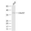
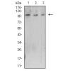
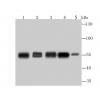
Fig1: Western blot analysis of PARP1 on different lysates using anti-PARP1 antibody at 1/100 dilution.
Positive control:
Lane 1: Daudi
Lane 2: Rat spleen tissue
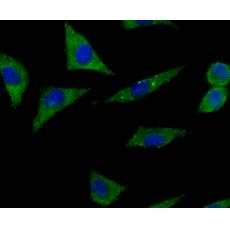
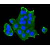
Fig2: ICC staining PARP1 (green) in 293T cells. The nuclear counter stain is DAPI (blue). Cells were fixed in paraformaldehyde, permeabilised with 0.25% Triton X100/PBS.
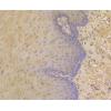
Fig3: Immunohistochemical analysis of paraffin-embedded human tonsil tissue using anti- PARP1 antibody. Counter stained with hematoxylin.
Fig4: Immunohistochemical analysis of paraffin-embedded human pancreas tissue using anti- PARP1 antibody. Counter stained with hematoxylin.
Fig5: Immunohistochemical analysis of paraffin-embedded rat brain tissue using anti- PARP1 antibody. Counter stained with hematoxylin.
Fig6: Immunohistochemical analysis of paraffin-embedded mouse testis tissue using anti- PARP1 antibody. Counter stained with hematoxylin.
Fig7: Flow cytometric analysis of Daudi cells with PARP1 antibody at 1/100 dilution (red) compared with an unlabelled control (cells without incubation with primary antibody; black).
特别提示:本公司的所有产品仅可用于科研实验,严禁用于临床医疗及其他非科研用途!